. 2023 Jul;152(1):94-106.e12.
doi: 10.1016/j.jaci.2023.02.028. Epub 2023 Mar 8.

CCL5 is a potential bridge between type 1 and type 2 inflammation in asthma

Affiliations

CCL5 is a potential bridge between type 1 and type 2 inflammation in asthma

Marc Gauthier et al. J Allergy Clin Immunol. 2023 Jul.

Abstract

Background: Type 1 (T1) inflammation (marked by IFN-γ expression) is now consistently identified in subsets of asthma cohorts, but how it contributes to disease remains unclear.

Objective: We sought to understand the role of CCL5 in asthmatic T1 inflammation and how it interacts with both T1 and type 2 (T2) inflammation.

Methods: CCL5, CXCL9, and CXCL10 messenger RNA expression from sputum bulk RNA sequencing, as well as clinical and inflammatory data were obtained from the Severe Asthma Research Program III (SARP III). CCL5 and IFNG expression from bronchoalveolar lavage cell bulk RNA sequencing was obtained from the Immune Mechanisms in Severe Asthma (IMSA) cohort and expression related to previously identified immune cell profiles. The role of CCL5 in tissue-resident memory T-cell (TRM) reactivation was evaluated in a T1high murine severe asthma model.

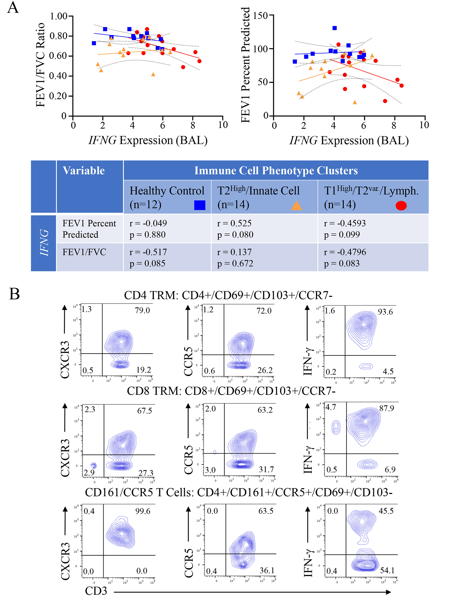
Results: Sputum CCL5 expression strongly correlated with T1 chemokines (P < .001 for CXCL9 and CXCL10), consistent with a role in T1 inflammation. CCL5high participants had greater fractional exhaled nitric oxide (P = .009), blood eosinophils (P < .001), and sputum eosinophils (P = .001) in addition to sputum neutrophils (P = .001). Increased CCL5 bronchoalveolar lavage expression was unique to a previously described T1high/T2variable/lymphocytic patient group in the IMSA cohort, with IFNG trending with worsening lung obstruction only in this group (P = .083). In a murine model, high expression of the CCL5 receptor CCR5 was observed in TRMs and was consistent with a T1 signature. A role for CCL5 in TRM activation was supported by the ability of the CCR5 inhibitor maraviroc to blunt reactivation.

Conclusion: CCL5 appears to contribute to TRM-related T1 neutrophilic inflammation in asthma while paradoxically also correlating with T2 inflammation and with sputum eosinophilia.

Keywords: Asthma; CCL5; CCR5; CXCL-10; CXCL9; IFN-γ; maraviroc; tissue-resident memory T cells (TRM).


Figures

Figure 1:
Figure 1:. CCL5 Expression in sputum correlates with T1 chemokines and is associated with multiple sputum cell differentials.
Bulk RNA sequencing data was obtained from sputum samples in the SARP III cohort at their baseline visit. (A) The expression levels of CCL5, CXCL9 and CXCL10 in sputum (median with inter-quartile range) are shown across the cohort by asthma severity (Healthy Control [Healthy], Mild to Moderate Asthma [MMA] and Severe Asthma [SA]); CCL5 High status was determined by using a cutoff of 1 SD above the healthy control median (y=9.185, dashed line); (B) CCL5 expression in sputum correlates with the expression of T1 chemokine CXCL10, Spearman non-parametric correlations, simple linear regression (black) with 95% CI (shaded blue); (C) Sputum cell differential and count for lymphocytes shown by CCL5 High/Low status, Mann-Whitney U (D) FEV1 % predicted and FEV1/FVC ratio shown by CCL5 High/Low status, linear regression adjusted for age and BMI.
Figure 2:
Figure 2:. CCL5 High Status is associated with clinical T2 biomarkers and both eosinophilic and neutrophilic sputum:
(A) Absolute blood eosinophil counts and fraction of exhaled nitric oxide (FeNO) are compared by CCL5 group, Mann-Whitney U test (B) Sputum cell differential for eosinophils shown by CCL5 High/Low status, Mann-Whitney U (C) Sputum cell differential for neutrophils shown by CCL5 High/Low status, Mann-Whitney U (D) Sputum eosinophilia (defined by >2% eosinophils), sputum neutrophilia (defined as above the median value) and mixed eosinophilic/neutrophilic (meeting both criteria) are tabulated against CCL5 High/Low status, values shown as percentages of the whole CCL5 group, Fisher’s exact test calculated using contingency tables based on subject numbers.
Figure 3:
Figure 3:. IFNG and CCL5 Expression differ among BAL inflammatory cellular profiles with differing clinical features.
(A) IFNG and CCL5 expression in BAL by bulk RNA sequencing show no significant differences when grouped by asthma severity, however when grouped by previously identified BAL cell profiles significant elevation is seen in the lymphocytic dominant group, Kruskal-Wallis with Dunn’s post-hoc testing when significant, exact p-values shown (B) Correlation of CCL5 expression with IFNG (T1 cytokine) and IL4 (T2 cytokine) (C) Correlation of CCL5 with Eosinophil percentage in blood as well as BAL; Spearman’s rho, simple linear regression (black) with 95% CI (shaded blue).
Figure 4:
Figure 4:. Associations of IFNG expression with measures of obstruction by inflammatory group and TRM receptor and cytokine expression:
(A) BAL IFNG expression was correlated with forced expiratory volume at 1 second (FEV1) percent predicted and FEV1/Forced Vital Capacity (FVC) by BAL inflammatory cell phenotype group (Healthy Control, T2High/Innate Cell, T1High/T2Var./Lymphocyte). Spearman non-parametric correlation for each comparison by group are shown in the table below (B) Human BAL CD4 and CD8 TRMs, and CD4 CD161+/CCR5+ cells identified by CyTOF and machine learning with the R implementation of FlowSOM (see Fig E1) were assessed for expression of CXCR3, CCR5, and IFN-γ using conventional analysis via FlowJo software; representative plots from 1 participant shown.
Figure 5
Figure 5. Identification of Tissue Resident Memory T-cells and CD161+CCR5+ Cells in a Murine T1 severe asthma model:
(A) Cells isolated from the lung were stained and examined by flow cytometry; Gating on CD3+CD4+ (CD4) and CD3+CD8+ (CD8) cells, tissue resident memory T-cells (TRMs) were identified by expression of CD69+CD103+ with numbers of both CD4 and CD8 TRMs significantly expressed in the severe asthma model (SA) compared to naïve mice (Mann-Whitney U-test, data representative of two independent experiments) (B) Examination of CD4 and CD8 TRMs for surface staining for CXCR3 and CCR5 identified an abundance of CXCR3+ TRMs co-expressing CCR5 (C) Gating on CD161, a population of CD161+CCR5+ cells was identified (data pooled from 4 mice per group) (D/E) Mice underwent treatment with either the CCR5 inhibitor maraviroc (300mg/1L drinking water) or vehicle (water) beginning during the rest phase of the model (Fig E2); following reactivation challenge, cells were isolated from the lungs and underwent intracellular and surface staining for total lung cells and then gating on TRMs for IFN-γ+ and IL-17+ cells along with CXCR3+CCR5+ cells (E) (data pooled from two separate experiments, Student’s T-test with Welch’s correction).

References

    1. Rose D, Mannino DM, Leaderer BP. Asthma Prevalence Among US Adults, 1998–2000: Role of Puerto Rican Ethnicity and Behavioral and Geographic Factors. American Journal of Public Health 2006; 96:880–8. - PMC - PubMed
    1. Hekking PP, Wener RR, Amelink M, Zwinderman AH, Bouvy ML, Bel EH. The prevalence of severe refractory asthma. J Allergy Clin Immunol 2015; 135:896–902. - PubMed
    1. Gauthier M, Ray A, Wenzel SE. Evolving Concepts of Asthma. Am J Respir Crit Care Med 2015; 192:660–8. - PMC - PubMed
    1. Wenzel S Severe asthma: from characteristics to phenotypes to endotypes. Clin Exp Allergy 2012; 42:650–8. - PubMed
    1. Brusselle GG, Koppelman GH. Biologic Therapies for Severe Asthma. New England Journal of Medicine 2022; 386:157–71. - PubMed

Publication types