Review
. 2023 Apr:244:108372.
doi: 10.1016/j.pharmthera.2023.108372. Epub 2023 Mar 7.

Endotheliopathy in the metabolic syndrome: Mechanisms and clinical implications

Affiliations
Review

Endotheliopathy in the metabolic syndrome: Mechanisms and clinical implications

Kunimaro Furuta et al. Pharmacol Ther. 2023 Apr.

Abstract

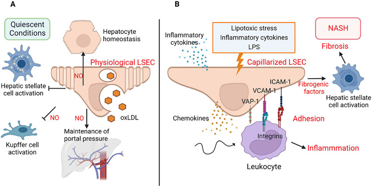
The increasing prevalence of the metabolic syndrome (MetS) is a threat to global public health due to its lethal complications. Nonalcoholic fatty liver disease (NAFLD) is the hepatic manifestation of the MetS characterized by hepatic steatosis, which is potentially progressive to the inflammatory and fibrotic nonalcoholic steatohepatitis (NASH). The adipose tissue (AT) is also a major metabolic organ responsible for the regulation of whole-body energy homeostasis, and thereby highly involved in the pathogenesis of the MetS. Recent studies suggest that endothelial cells (ECs) in the liver and AT are not just inert conduits but also crucial mediators in various biological processes via the interaction with other cell types in the microenvironment both under physiological and pathological conditions. Herein, we highlight the current knowledge of the role of the specialized liver sinusoidal endothelial cells (LSECs) in NAFLD pathophysiology. Next, we discuss the processes through which AT EC dysfunction leads to MetS progression, with a focus on inflammation and angiogenesis in the AT as well as on endothelial-to-mesenchymal transition of AT-ECs. In addition, we touch upon the function of ECs residing in other metabolic organs including the pancreatic islet and the gut, the dysregulation of which may also contribute to the MetS. Finally, we highlight potential EC-based therapeutic targets for human MetS, and NASH based on recent achievements in basic and clinical research and discuss how to approach unsolved problems in the field.

Keywords: Adipose tissue (AT); Capillarization; Endothelial cell (EC); Endothelial-to-mesenchymal transition (EndoMT); Endotheliopathy; Fibrosis; Inflammation; Liver sinusoidal endothelial cell (LSEC); Metabolic syndrome (MetS); Nonalcoholic fatty liver disease (NAFLD); Nonalcoholic steatohepatitis (NASH).


Conflict of interest statement

Declaration of Competing Interest The authors have declared that no conflict of interest exists.

Figures

Figure 1.
Figure 1.. The role of LSECs under quiescent conditions and in NASH
(A) In quiescent conditions, LSEC endocytosis facilitates the transfer of oxidized LDL from the circulation to the hepatocytes, thereby maintaining systemic lipid homeostasis. In addition, LSEC intrinsically exhibit anti-inflammatory and anti-fibrotic roles through inhibiting the activation of Kupffer cells and hepatic stellate cells (HSCs), respectively. LSEC-derived NO contributes to the lipid homeostasis in hepatocytes by driving β-oxidation in mitochondria. Furthermore, LSECs in physiological condition can regulate vascular resistance and portal pressure. (B) In NASH, with the stimulation of lipotoxic stress, proinflammatory cytokines and gut bacteria-derived lipopolysaccharide, LSECs acquire a pro-inflammatory and pro-fibrogenic phenotype, and become capillarized by losing their fenestrae and gaining a basement membrane a process called endotheliopathy. Capillarized LSECs release pro-inflammatory mediators including cytokines and chemokines and express luminal adhesion molecules promoting leukocyte adhesion to LSECs via their cognate binding integrin receptors. In addition, capillarized LSECs secrete fibrogenic factors and drive HSC activation, thereby promoting hepatic fibrosis. NO, nitric oxide; oxLDL, oxidized low density lipoprotein; LPS, lipopolysaccharide; VCAM-1, vascular cell adhesion molecule 1; ICAM-1, intracellular adhesion molecule 1; VAP-1, vascular adhesion protein 1.
Figure 2.
Figure 2.. Molecular mediators of EC crosstalk in adipose tissue in health and metabolic syndrome.
(A) In healthy condition, quiescent ECs maintain the capillary integrity and release NO and EVs, while adipocytes produce adiponectin, all of which promote AT homeostasis. (B) In MetS, adipocytes secrete more pro-inflammatory factors, such as TNFα, IL-6, IL-1β, leptin and less adiponectin, leading to increase immune infiltration. Meanwhile, ECs secrete pro-inflammatory signals such as ROS, ET-1, TNFα and TSP1, and reduce NO production, leading to increased vascular permeability. The expanding adipocytes cause tissue hypoxia and increase VEGF production, switching ECs from quiescent to proliferative and angiogenic state. However, severe obesity or advanced MetS causes vascular rarefaction, chronic inflammation and likely EndoMT. The latter may be induced by TGF-β and leads to release of more EVs to induce EC dysfunction. TSP1, Thrombospondin-1; VEGF, vascular endothelial growth factor; EVs, extracellular vesicles; ET-1, endothelin-1; NO, nitric oxide; ROS, reactive oxygen species; TNFα, tumor necrosis factor α, IL, interleukin; MCP1, monocyte chemoattractant protein-1.
Figure 3.
Figure 3.. Mechanisms of ECs residing in pancreatic islet and the gut in MetS development.
In a healthy state, there is an active exchange of factors between endocrine cells, e.g., islet β-cells and ECs in the islets and between intestinal epithelial cells with ECs in the gut. VEGF and INS are secreted from β cells to modulate EC function. Reciprocally, ECs can secrete HGF to stimulate β cell proliferation. In the gut, ECs secrete caspase-8 to maintain gut homeostasis. GIP is secreted from the intestines in response to food intake. The intestine also produces GLP-1, which enters the circulation and is up taken by the pancreas where it promotes insulin secretion. The gut microbiota also secretes metabolites (blue spheres) to help maintain the homeostasis of the gut and whole-body metabolism. During the development of MetS, the gut bacteria can travel through the epithelial layer and into the vasculature, where it secretes LPS to activate ECs. The intestinal epithelial cells can also produce TNFα and IL-1β, contributing to EC dysfunction. Dysfunctional ECs can recruit monocytes for inflammatory activation in the pancreas and gut. The activated and dysfunctional ECs may secrete EVs to mediate intercellular and inter-organ crosstalk. VEGF, vascular endothelial growth factor; INS, insulin; HGF, hepatocyte growth factor; GLP-1, glucagon-like peptide-1; GIP, glucose-dependent insulinotropic polypeptide; LPS, lipopolysaccharide; VCAM-1, vascular cell adhesion molecule 1; ICAM-1, intracellular adhesion molecule 1; TNF-α, tumor necrosis factor-alpha; IL-1β, interleukin 1 beta; EVs, extracellular vesicles.

References

    1. Abe M, Matsuda M, Kobayashi H, Miyata Y, Nakayama Y, Komuro R, Fukuhara A, & Shimomura I (2008). Effects of statins on adipose tissue inflammation: their inhibitory effect on MyD88-independent IRF3/IFN-beta pathway in macrophages. Arterioscler Thromb Vasc Biol, 28, 871–877. - PubMed
    1. Adams DH, Hubscher SG, Fisher NC, Williams A, & Robinson M (1996). Expression of E-selectin and E-selectin ligands in human liver inflammation. Hepatology, 24, 533–538. - PubMed
    1. Aird WC (2004). Endothelium as an organ system. Crlt Care Med, 32, S271–279. - PubMed
    1. Aird WC (2007). Phenotypic heterogeneity of the endothelium: II. Representative vascular beds. Circ Res, 100, 174–190. - PubMed
    1. Aizarani N, Saviano A, Sagar, Mailly L, Durand S, Herman JS, Pessaux P, Baumert TF, & Grun D (2019). A human liver cell atlas reveals heterogeneity and epithelial progenitors. Nature, 572, 199–204. - PMC - PubMed

Publication types

MeSH terms