. 2023 Aug;76(8):561-565.
doi: 10.1136/jcp-2023-208771. Epub 2023 Mar 9.

Identification of a protein expression signature distinguishing early from organising diffuse alveolar damage in COVID-19 patients

Affiliations

Identification of a protein expression signature distinguishing early from organising diffuse alveolar damage in COVID-19 patients

Helen Ashwin et al. J Clin Pathol. 2023 Aug.

Abstract

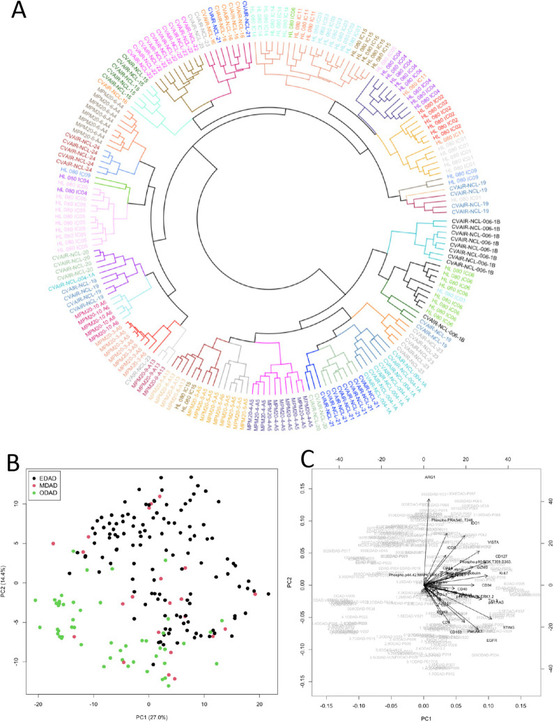
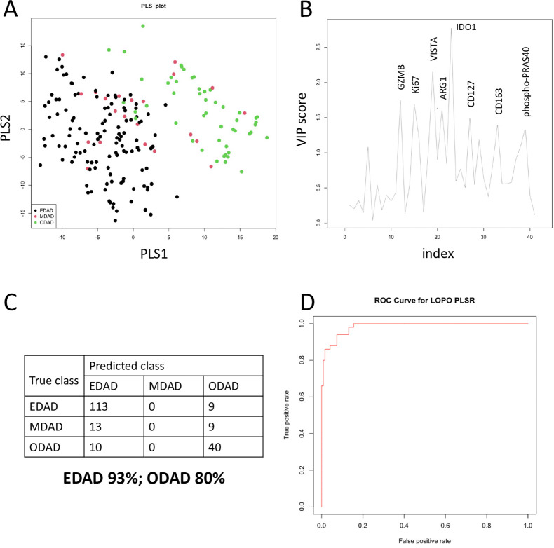
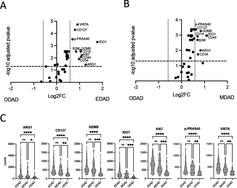
Diffuse alveolar damage (DAD) is the histological expression of acute respiratory distress syndrome and characterises lung pathology due to infection with SARS-CoV-2, and other respiratory pathogens of clinical significance. DAD reflects a time-dependent immunopathological process, progressing from an early/exudative stage through to an organising/fibrotic stage, yet within an individual these different stages of DAD may coexist. Understanding the progression of DAD is central to the development of new therapeutics to limit progressive lung damage. Here, we applied highly multiplexed spatial protein profiling to autopsy lung tissues derived from 27 patients who died from COVID-19 and identified a protein signature (ARG1, CD127, GZMB, IDO1, Ki67, phospho-PRAS40 (T246) and VISTA) that distinguishes early DAD from late DAD with good predictive accuracy. These proteins warrant further investigation as potential regulators of DAD progression.

Keywords: COVID-19; INFLAMMATION; Immune System Diseases; LUNG.


Conflict of interest statement

Competing interests: None declared.

Figures

Figure 1
Figure 1
Analysis of protein DSP data. (A) Circular dendrogram from hierarchical clustering of protein DSP ROIs (with patient identifiers colour-coded and clusters coloured separately; see online supplemental table S1. (B, C) Principal component analysis (PCA) scores plot for the first two principal components coloured by EDAD, MDAD and ODAD (B) and with loadings shown as vectors (C). DSP, digital spatial profiling; EDAD, exudative DAD; MDAD, mixed DAD; ODAD, organising DAD; ROI, regions of interest.
Figure 2
Figure 2
Discrimination of DAD classes based on protein signature. (A, B) Partial least squares analysis of EDAD, MDAD and ODAD samples shown as PLS plot (A) and by variable importance in projection (VIP) score (B). (C) Confusion matrix for results of PLS-LDA leave one patient out prediction using eight variables with VIP scores >1.3 (GZMB, Ki.67, VISTA, ARG1, IDO1, CD127, CD163, Phospho.PRAS40). (D) Receiver operating characteristic (ROC) curve generated for EDAD versus ODAD ROIs. DAD, diffuse alveolar damage; EDAD, exudative DAD; LDA, linear discriminate analysis; MDAD, mixed DAD; ODAD, organising DAD; PLS, partial least square; ROI, regions of interest.
Figure 3
Figure 3
Differential target expression between EDAD and ODAD using linear mixed modelling. (A, B) Differentially expressed (FDR 5%; FC=1.5) protein targets between EDAD and ODAD (A) and MDAD and ODAD (B). Data derives from a linear mixed modelling with patient repeat measures and cohort as a random effect. (C) Individual ROI counts for EDAD, MDAD and ODAD ROIs for identified target proteins. *p<0.05, **p<0.01, ***p<0.001, ****p<0.0001 between indicated groups. ns, non-significant; EDAD, exudative DAD; MDAD, mixed DAD; ODAD, organising DAD; ROIs, regions of interest. FDR, false discovery rate.

References

    1. Centre JHCR . Available: https://coronavirus.jhu.edu/map.html
    1. Li Y, Wu J, Wang S, et al. Progression to fibrosing diffuse alveolar damage in a series of 30 minimally invasive autopsies with COVID-19 pneumonia in Wuhan, China. Histopathology 2021;78:542–55. 10.1111/his.14249 - DOI - PMC - PubMed
    1. Milross L, Majo J, Cooper N, et al. Post-mortem lung tissue: the fossil record of the pathophysiology and immunopathology of severe COVID-19. Lancet Respir Med 2022;10:95–106. 10.1016/S2213-2600(21)00408-2 - DOI - PMC - PubMed
    1. Nienhold R, Ciani Y, Koelzer VH, et al. Two distinct immunopathological profiles in autopsy lungs of COVID-19. Nat Commun 2020;11:5086. 10.1038/s41467-020-18854-2 - DOI - PMC - PubMed
    1. Damiani S, Fiorentino M, De Palma A, et al. Pathological post-mortem findings in lungs infected with SARS-cov-2. J Pathol 2021;253:31–40. 10.1002/path.5549 - DOI - PubMed