. 2023 Mar 10;6(1):254.
doi: 10.1038/s42003-023-04609-4.

YgfB increases β-lactam resistance in Pseudomonas aeruginosa by counteracting AlpA-mediated ampDh3 expression

Affiliations

YgfB increases β-lactam resistance in Pseudomonas aeruginosa by counteracting AlpA-mediated ampDh3 expression

Ole Eggers et al. Commun Biol. .

Abstract

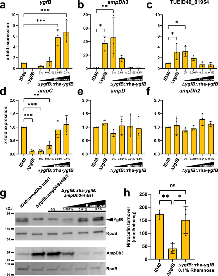
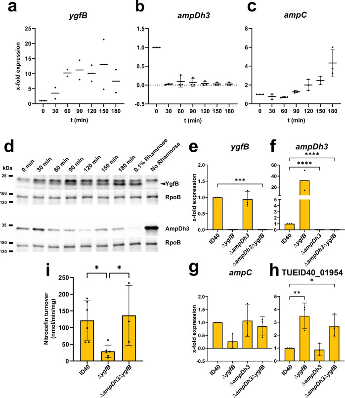
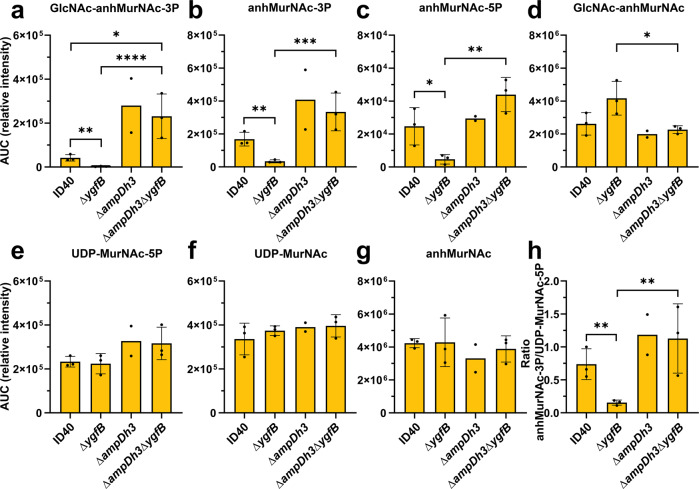
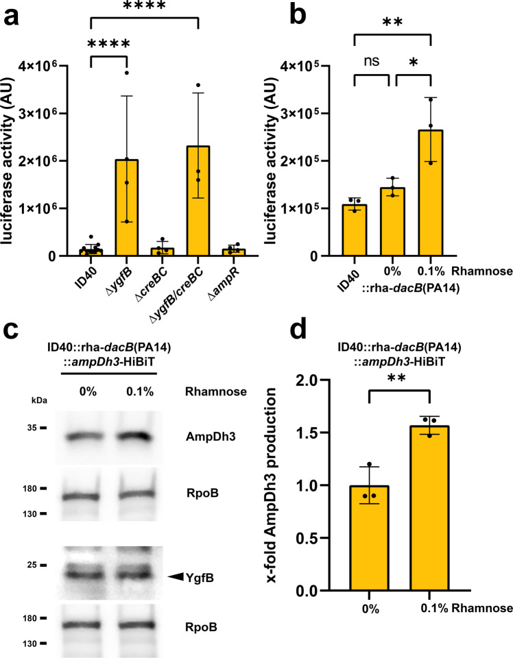
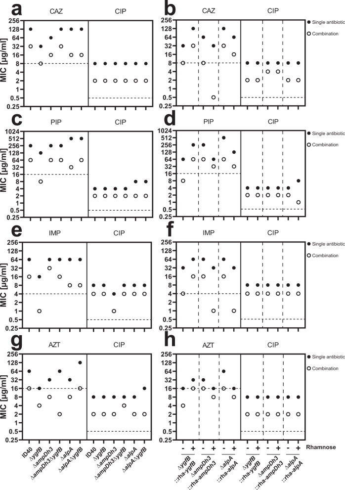
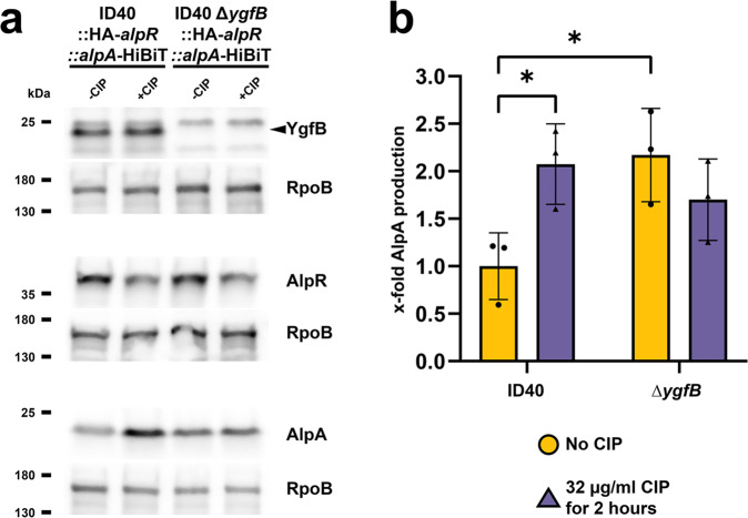
YgfB-mediated β-lactam resistance was recently identified in multi drug resistant Pseudomonas aeruginosa. We show that YgfB upregulates expression of the β-lactamase AmpC by repressing the function of the regulator of the programmed cell death pathway AlpA. In response to DNA damage, the antiterminator AlpA induces expression of the alpBCDE autolysis genes and of the peptidoglycan amidase AmpDh3. YgfB interacts with AlpA and represses the ampDh3 expression. Thus, YgfB indirectly prevents AmpDh3 from reducing the levels of cell wall-derived 1,6-anhydro-N-acetylmuramyl-peptides, required to induce the transcriptional activator AmpR in promoting the ampC expression and β-lactam resistance. Ciprofloxacin-mediated DNA damage induces AlpA-dependent production of AmpDh3 as previously shown, which should reduce β-lactam resistance. YgfB, however, counteracts the β-lactam enhancing activity of ciprofloxacin by repressing ampDh3 expression and lowering the benefits of this drug combination. Altogether, YgfB represents an additional player in the complex regulatory network of AmpC regulation.


Conflict of interest statement

The authors declare no competing interest.

Figures

Fig. 1
Fig. 1. Validation of transcriptomic data.
RNA was isolated from the indicated strains and RT-qPCR performed. Data depict the mean and SD of the x-fold difference in mRNA expression compared to ID40 of (a) ygfB, (b) ampDh3, (c) TUEID40_01954, (d) ampC, (e) ampD, and (f) ampDh2 for n = 3 individual experiments. Asterisks depict significant differences (*p < 0.05, **p < 0.01, ****p < 0.001; one-way ANOVA, Dunnett’s multiple comparison comparing to ID40). g Whole cell lysates of the indicated strains were used for SDS-PAGE and Western blots. The detection of YgfB and AmpDh3 was done on separate Western blots, each with RpoB as a loading control. As primary antibodies, anti-YgfB or as a loading control anti-RpoB antibodies and as secondary antibody anti-IgG-HRP antibodies were used and detection was done using ECL. For determination of AmpDh3, recombinant LgBiT was used. LgBiT binds to HiBiT resulting in a functional luciferase. The cleavage of the substrate furimazine leads to detectable chemiluminescence. Data are representative of three independent experiments. h Whole cell lysates from the indicated strains were used to determine β-lactamase activity using a nitrocefin assay. Data depict the mean and SD of nitrocefin turnover for n = 3 individual experiments. Asterisks depict significant differences (ns p > 0.05, *p < 0.05, **p < 0.01; one-way ANOVA, Tukey’s multiple comparisons).
Fig. 2
Fig. 2. Relationship between YgfB, AmpDh3 and AmpC.
The expression of ygfB in ID40∆ygfB::rha-ygfB was induced with 0.1% rhamnose at time point zero and RNA was isolated at various time points of growth in LB medium for further use in RT-qPCRs. mRNA expression of (a) ygfB, (b) ampDh3 and (c) ampC was measured. Data depict the mean and SD of x-fold expression compared to ID40 0 min of n = 2–3 independent experiments. d The expression of ygfB in ID40∆ygfB::rha-ygfB::ampDh3-HiBiT was induced with 0.1% rhamnose at time point zero and samples were taken from the growing culture in LB medium at the indicated time points. Then, whole cell lysates were prepared and used for SDS-PAGE and Western blots. The 0.1% condition depicts a strain grown under constant rhamnose supplementation. The detection of YgfB and AmpDh3 was done on separate Western blots, each with RpoB as a loading control. As primary antibodies, anti-YgfB or anti-RpoB antibodies and as secondary antibody anti-IgG-HRP antibodies were used and detection was done using ECL. For determination of AmpDh3, recombinant LgBiT was used. LgBiT binds to HiBiT resulting in a functional luciferase. The cleavage of the substrate furimazine leads to detectable chemiluminescence. Data are representative of three independent experiments. eh P. aeruginosa strains as indicated were grown for 3 h in LB. RNA was isolated and used for RT-qPCR. mRNA expression of (e) ygfB, (f) ampDh3, (g) ampC and (h)TUEID40_01954 was measured. Data depict mean and SD of x-fold mRNA expression compared to ID40 of n = 2–3 independent experiments. i β-lactamase activity of indicated strains was determined by using a nitrocefin assay. Data depict mean and SD of n = 3–6 independent experiments. An asterisks indicate significant differences compared to ID40 (*p < 0.05, **p < 0.01, ***p < 0.001, ****p < 0.0001; one-way ANOVA, (eh) Dunnett’s multiple comparisons comparing to ID40, (i) Šídák’s multiple comparisons).
Fig. 3
Fig. 3. Impact of YgfB and AmpDh3 on the composition of peptidoglycan precursors.
The indicated P. aeruginosa strains were grown for 6 h in LB medium, cytosolic extracts were generated and analyzed by HPLC-mass spectrometry. The graphs depict the mean and SD of the area under curve (AUC) of the peaks obtained for (a) GlcNAc-anhMurNAc-3P, (b) anhMurNAc-3P, (c) anhMurNAc-5P, (d) GlcNAc-anhMurNAc, (e) UDP-MurNAc-5P, (f) UDP-MurNAc and (g) anhMurNAc of n = 2–3 independent experiments. In addition, in (h) the ratio between anhMurNAc-3P and UDP-MurNAc-5P is shown. An asterisks indicate significant differences (*p < 0.05, **p < 0.01, ***p < 0.001, ****p < 0.0001); Values were log10 transformed, logarithmic values were shown to be normally distributed according to Wilks-test, one-way ANOVA of log10 transformed values, Tukey’s multiple comparisons.
Fig. 4
Fig. 4. Analysis of the ampDh3 promoter.
a Schematic view of the putative ampDh3 promoter. Numbers below depict the base pairs counted upstream of the coding sequence (CDS). The black bar in which blue AlpA is depicted marks the AlpA binding element (ABE) as defined by Peña et al.. Stop symbols mark terminator regions as predicted. b, c Luciferase assays were performed to measure transcriptional activity using ampDh3 promoter fragments of various lengths fused to the coding sequence of NanoLuc. Data depict the mean and SD of the x-fold luciferase activity of 0-luc compared to all other fragments of n = 3, n = 6 for (b) and n = 4 for (c) independent experiments. In (b), the strain ID40ΔygfB::rha-ygfB without (yellow circle, depicted as -YgfB) or with 0.1% rhamnose (purple triangle, depicted as +YgfB) and in (c) the strains ID40 (purple up-pointing triangle), ΔygfB (yellow dot), ΔalpA (white diamond) and ΔygfBΔalpA (mint down-pointing triangle) were used. The strains carry the indicated pBBR plasmids in which the expression of NanoLuc is under the control of fragments of different length of the ampDh3 promoter. For instance, 532 is the abbreviation for a pBBR plasmid containing an ampDh3 promoter fragment comprising the region –532 to –1 bp upstream of the CDS of NanoLuc. In (d) the strains ∆alpA::rha-alpA and ∆ygfBalpA::rha-alpA carrying pBBR-532-luc were used as indicated. Data depict the mean and SD of the x-fold luciferase activity compared to ∆alpA::rha-alpA without rhamnose added for n = 3 replicates. Asterisks in (d) indicate significant differences (****p < 0.0001; one-way ANOVA, Šídák’s multiple comparisons).
Fig. 5
Fig. 5. Interrelationship between creBC, dacB and ampDh3.
a, b Luciferase reporter assays using the indicated strains harboring the reporter construct ampDh3-532-luc comprising the ampDh3 promoter fragment between position -532 and -1 upstream of the CDS of Nanoluc. Data depict the mean and SD of luciferase activity of n = 12, 4, 4, 3, 4 for (a) and for (b) n = 3 independent experiments. Asterisks in (a) indicate significant differences compared to ID40 (****p < 0.0001; one-way ANOVA, Dunnett’s multiple comparisons comparing to ID40) and in (b) compared between all conditions (ns p > 0.05, *p < 0.05, **p < 0.01; one-way ANOVA, Tukey’s multiple comparisons). c ID40::rha-dacB(PA14)::ampDh3-HiBiT was grown for 3 h without or with 0.1% rhamnose to induce the expression of dacB of PA14 in LB medium at 37 °C. Whole cell lysates were used to perform SDS-PAGE and Western blot transfer. The detection of YgfB and AmpDh3 was done on separate Western blots, each with RpoB as a loading control. As primary antibodies anti-YgfB or anti-RpoB antibodies and as secondary antibodies anti-IgG-HRP antibodies were used and detection was done using ECL. AmpDh3-HiBiT was detected by incubating the membrane with LgBiT and furimazine. Data are representative for three independent experiments. d Bacteria were harvested, lysed and subsequently luciferase activity of AmpDh3-HiBiT was measured as described in the “Methods” section. Data depict the mean and SD of the x-fold luciferase activity compared to 0% rhamnose for n = 3 independent experiments. Asterisks indicate significant differences (p** < 0.01; unpaired, two-tailed t test).
Fig. 6
Fig. 6. Checkerboard assays to investigate combinatory effects of ciprofloxacin and β-lactams.
The indicated antibiotics were combined in log2-fold dilutions and minimum inhibitory concentrations for antibiotics alone or in combination were determined. Plotted are the MICs for single antibiotics (filled circle) and combination of antibiotics (open circle). The figure shows the combination of CAZ and CIP, with the single mutants in (a) and conditional deletion mutants grown in the absence or presence of 0.1% rhamnose shown in (b). The combination of PIP and CIP, with the single mutants is shown in (c) and conditional deletion mutants grown in the absence or presence of 0.1% rhamnose shown in (d). The combination of IMP and CIP, with the single mutants is shown in (e) and conditional deletion mutants grown in the absence or presence of 0.1% rhamnose shown in (f). The combination of AZT and CIP, with the single mutants is shown in (g) and conditional deletion mutants grown in the absence or presence of 0.1% rhamnose shown in (h). Dotted horizontal lines show resistance break points for each antibiotic according to EUCAST. CAZ ceftazidime, PIP piperacillin, AZT aztreonam, IMP imipenem, CIP ciprofloxacin. Data are derived from two independent experiments for each combination.
Fig. 7
Fig. 7. Modulation of AlpR and AlpA production by YgfB and ciprofloxacin (CIP).
a Whole cell lysates of the indicated strains were used for Western blot analyses. +CIP conditions were treated with 32 µg/ml of ciprofloxacin for 2 h. The detection of YgfB, AlpR and AlpA was done on separate Western blots, each with RpoB as a loading control. As primary antibodies, anti-HA, anti-YgfB or anti-RpoB antibodies and as secondary antibody anti-IgG-HRP antibodies were used and detection was done using ECL. For detection of AlpA-HiBiT, recombinant LgBiT was used. LgBiT binds to HiBiT resulting in a functional luciferase. The cleavage of the substrate furimazine leads to detectable chemiluminescence. Data are representative of three independent experiments. b Quantification of AlpA by measuring luciferase activity of AlpA-HiBiT in lysed cell extracts. Conditions not treated with CIP depicted as yellow circle, conditions with added CIP depicted as purple up-pointing triangle. Data depict mean and SD of x-fold luciferase activity compared to ID40 wildtype for n = 3 independent experiments. An asterisks indicate significant differences compared to ID40 -CIP (*p < 0.05; two-way ANOVA, Šídák’s multiple comparisons comparing to ID40 -CIP).
Fig. 8
Fig. 8. Interaction of AlpA with YgfB.
Pulldowns were performed using (a) recombinant GST-YgfB or GST as a bait and cell lysates of ID40∆ygfB::alpA-HiBiT::HA-alpR as the prey and (b) recombinant His-MBP or His-MBP-AlpA as a bait and recombinant YgfB as the prey. Shown are Western blots using either LgBiT or anti-YgfB antibodies for detection. Of note: the slight difference in migration of AlpA/YgfB in input and eluate is associated with the different composition of input and elution buffer (a) is representative for five experiments, (b) for two experiments.
Fig. 9
Fig. 9. Ciprofloxacin-induced signaling pathway leading to modulation of ampC expression.
a Ciprofloxacin triggers DNA damage which promotes autocleavage of AlpR (yellow) and as a consequence, the derepression of the alpA promoter. AlpA (red) acts as an antiterminator of ampDh3 expression increasing AmpDh3 (blue) production. AmpDh3 cleaves anhMurNAc-peptides (black triangle) which changes the balance of the AmpR-activator upon binding anhMurNAc-peptides and the AmpR-repressor upon binding UDP-MurNAc-5P (black square) in favor of AmpR-mediated repression of its target promoters (AmpR depicted as green). This leads to reduced ampC expression (purple) and decreased β-lactam resistance. b YgfB directly interacts with AlpA and blocks binding of AlpA to the ampDh3 promoter. Thereby, AlpA’s antiterminator function is inhibited and ampDh3 expression dampened. Reduced levels of AmpDh3 in the cytosol change the balance of AmpR-activating anhMurNAc-peptides and AmpR-repressing UDP-MurNAc-5P in favor of AmpR-mediated activation of its target promoters. This enhances AmpC production and leads to increased β-lactam resistance.

References

    1. Yoshimura F, Nikaido H. Permeability of Pseudomonas aeruginosa outer membrane to hydrophilic solutes. J. Bacteriol. 1982;152:636–642. doi: 10.1128/jb.152.2.636-642.1982. - DOI - PMC - PubMed
    1. Strateva T, Yordanov D. Pseudomonas aeruginosa—a phenomenon of bacterial resistance. J. Med. Microbiol. 2009;58:1133–1148. doi: 10.1099/jmm.0.009142-0. - DOI - PubMed
    1. Schmidtke AJ, Hanson ND. Role of ampD homologs in overproduction of AmpC in clinical isolates of Pseudomonas aeruginosa. Antimicrob. Agents Chemother. 2008;52:3922–3927. doi: 10.1128/AAC.00341-08. - DOI - PMC - PubMed
    1. Tam VH, et al. Prevalence of AmpC over-expression in bloodstream isolates of Pseudomonas aeruginosa. Clin. Microbiol Infect. 2007;13:413–418. doi: 10.1111/j.1469-0691.2006.01674.x. - DOI - PubMed
    1. Moya B, et al. Beta-lactam resistance response triggered by inactivation of a nonessential penicillin-binding protein. PLoS Pathog. 2009;5:e1000353. doi: 10.1371/journal.ppat.1000353. - DOI - PMC - PubMed

Publication types

MeSH terms