. 2023 Mar 9;3(1):19.
doi: 10.1038/s43705-023-00220-0.

A meta-analysis of the stony coral tissue loss disease microbiome finds key bacteria in unaffected and lesion tissue in diseased colonies

Affiliations

A meta-analysis of the stony coral tissue loss disease microbiome finds key bacteria in unaffected and lesion tissue in diseased colonies

Stephanie M Rosales et al. ISME Commun. .

Abstract

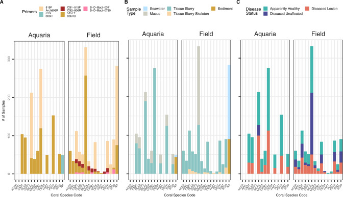
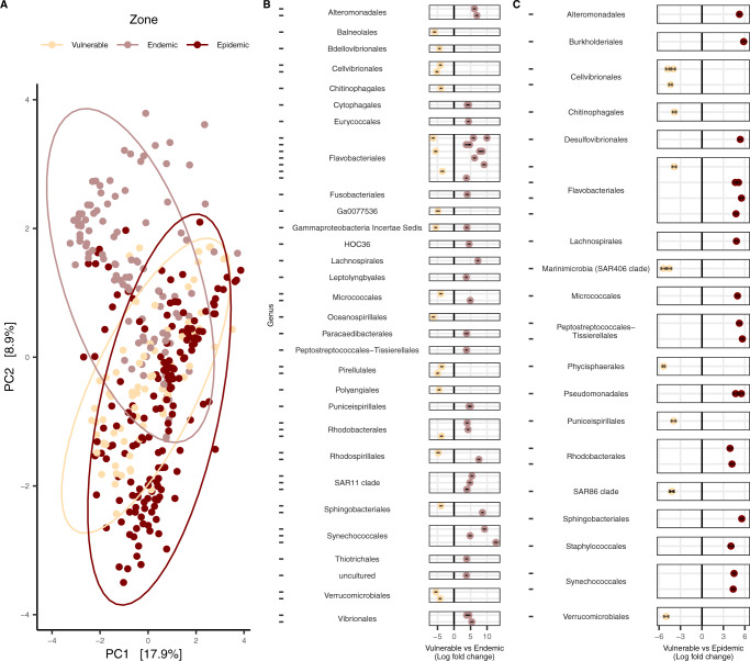
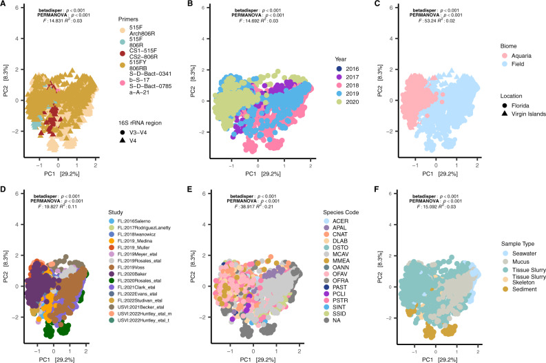
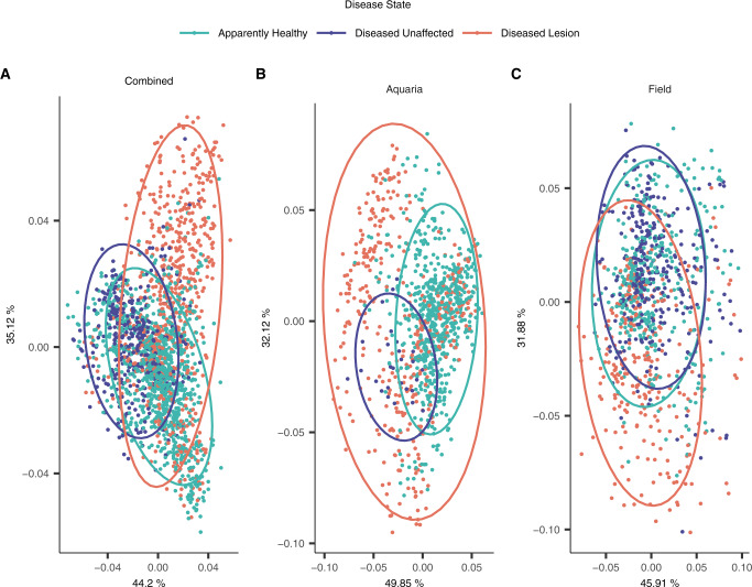
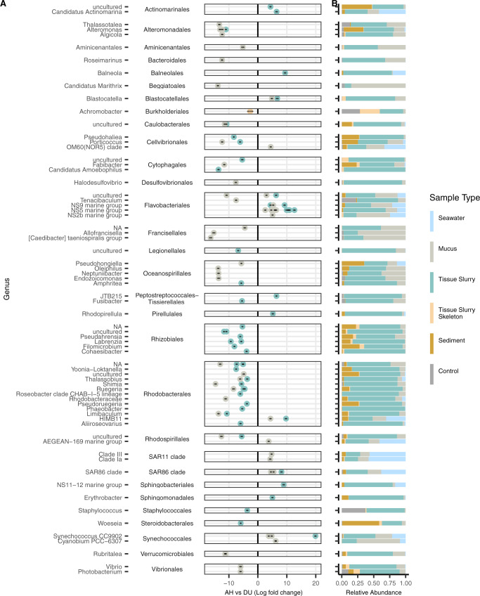
Stony coral tissue loss disease (SCTLD) has been causing significant whole colony mortality on reefs in Florida and the Caribbean. The cause of SCTLD remains unknown, with the limited concurrence of SCTLD-associated bacteria among studies. We conducted a meta-analysis of 16S ribosomal RNA gene datasets generated by 16 field and laboratory SCTLD studies to find consistent bacteria associated with SCTLD across disease zones (vulnerable, endemic, and epidemic), coral species, coral compartments (mucus, tissue, and skeleton), and colony health states (apparently healthy colony tissue (AH), and unaffected (DU) and lesion (DL) tissue from diseased colonies). We also evaluated bacteria in seawater and sediment, which may be sources of SCTLD transmission. Although AH colonies in endemic and epidemic zones harbor bacteria associated with SCTLD lesions, and aquaria and field samples had distinct microbial compositions, there were still clear differences in the microbial composition among AH, DU, and DL in the combined dataset. Alpha-diversity between AH and DL was not different; however, DU showed increased alpha-diversity compared to AH, indicating that, prior to lesion formation, corals may undergo a disturbance to the microbiome. This disturbance may be driven by Flavobacteriales, which were especially enriched in DU. In DL, Rhodobacterales and Peptostreptococcales-Tissierellales were prominent in structuring microbial interactions. We also predict an enrichment of an alpha-toxin in DL samples which is typically found in Clostridia. We provide a consensus of SCTLD-associated bacteria prior to and during lesion formation and identify how these taxa vary across studies, coral species, coral compartments, seawater, and sediment.


Conflict of interest statement

The authors declare no competing interests.

Figures

Fig. 1
Fig. 1. The number of aquaria and field samples for each coral species.
A small subunit (SSU) rRNA gene primer sets, B sample type, and C disease state. NAs in (A, B) represent sediment and seawater samples. Coral species codes represent the following: Acropora cervicornis (ACER), Acropora palmata (APAL), Colpophyllia natans (CNAT), Diploria labyrinthiformis (DLAB), Dichocoenia stokesii (DSTO), Montastraea cavernosa (MCAV), Meandrina meandrites (MMEA), Orbicella annularis (OANN), Orbicella faveolata (OFAV), Orbicella franksi (OFRA), Porites astreoides (PAST), Pseudodiploria clivosa (PCLI), Pseudodiploria strigosa (PSTR), Stephanocoenia intersepta (SINT), and Siderastrea siderea (SSID).
Fig. 2
Fig. 2. Comparisons among microbial communities of field-sourced apparently healthy (AH) coral colonies across stony coral tissue loss disease (SCTLD) zones (vulnerable, endemic, and epidemic).
A beta-diversity (centered log-ratio transformed and plotted with a Euclidean distance), and differential abundance analysis in (B) vulnerable vs endemic zones, and C vulnerable vs epidemic zones. ASVs are grouped by genus (represented by dashes) on the y axis and then by order, and only ASVs with a Padj < 0.001, a W statistic >90, and a log-fold change < −2 and >2 were visualized. AH samples from the three coral compartments (mucus, tissue slurry, and tissue slurry skeleton) were included and Acropora spp. samples were excluded from the analysis. The ellipses in (A) represent the center of the Euclidean distance from the respective zone with a 95% confidence of the ellipses.
Fig. 3
Fig. 3. Microbial beta-diversity of all coral species (stony coral tissue loss disease [SCTLD]-susceptible corals and Acropora spp.) and sample types (coral, sediment, and seawater) show differences within and between microbial communities.
A small subunit (SSU) 16S rRNA gene primers, B year, C biome, D study, E coral species, and F sample type. All plots were centered log-ratio transformed and visualized with a Euclidean distance. The NAs in (E) represent sediment and seawater samples; coral species codes are defined in Fig. 1 legend.
Fig. 4
Fig. 4. Microbial differences in coral disease state among apparently healthy colonies (AH), and unaffected (DU) and lesion (DL) areas on diseased colonies in beta-diversity using a robust Aitchison Distance.
A both aquaria and field samples (“Combined”), and B aquaria and C field samples only. Samples from Acropora spp. were excluded and the three coral compartments (mucus, tissue slurry, and tissue slurry skeleton) were included in this analysis. The ellipses represent the center of the Euclidean distance from the respective disease state with a 95% confidence of the ellipses.
Fig. 5
Fig. 5. Microbial amplicon sequence variants (ASVs) associated with unaffected areas on diseased colonies (DU).
Differential abundances between (A) apparently healthy (AH) vs DU. The y axis depicts ASVs grouped by genus and then by order. Only ASVs with a Padj < 0.001, W statistic >90, and a log-fold change <−1.5 and >1.5 were visualized. Coral compartments (i.e., mucus, tissue slurry, and tissue slurry skeleton) were included and Acropora spp. were excluded from this analysis. B The relative abundance of taxa enriched in AH and DU by sample type, which includes laboratory controls (“Control”) encompassing field, lab, kit, and mock communities.
Fig. 6
Fig. 6. Microbial amplicon sequence variants (ASVs) associated with lesions on diseased colonies (DL).
Differential abundances between (A) apparently healthy (AH) vs DL. The y axis depicts ASVs grouped by genus and then by order. Only ASVs with a Padj < 0.001, W statistic >90, and a log-fold change <−1.5 and >1.5 were visualized. Coral compartments (i.e., mucus, tissue slurry, and tissue slurry skeleton) were included and Acropora spp. were excluded from this analysis. B The relative abundance of taxa enriched in AH and DL by sample type, which includes laboratory controls (“Control”) encompassing field, lab, kit, and mock communities.
Fig. 7
Fig. 7. Co-occurrence networks of bacteria from weighted correlation network analysis (WGCNA) modules (Supplementary Fig. 7) among apparently healthy colonies (AH), and unaffected (DU) and lesion (DL) areas on diseased colonies.
The nodes represent amplicon sequence variants (ASV), which are sized by the ASV’s correlation value to its respective module. A triangle and label of the bacteria order denote that a node is a “key player.” The width of the edges corresponds to centrality, with thicker edges representing higher centrality. Samples from the three coral compartments (i.e., mucus, tissue slurry, and tissue slurry skeleton) were included in the analysis, and Acropora spp. samples were excluded from this analysis.

References

    1. Landsberg JH, Kiryu Y, Peters EC, Wilson PW, Perry N, Waters Y, et al. Stony coral tissue loss disease in Florida is associated with disruption of host—Zooxanthellae physiology. Front Mar Sci. 2020;7:576013. doi: 10.3389/fmars.2020.576013.. - DOI
    1. Case definition: stony coral tissue loss disease (SCTLD). https://floridadep.gov/sites/default/files/Copy%20of%20StonyCoralTissueL.... Accessed 12 July, 2022.
    1. Precht WF, Gintert BE, Robbart ML, Fura R, van Woesik R. Unprecedented disease-related coral mortality in southeastern Florida. Sci Rep. 2016;6. 10.1038/srep31374. - PMC - PubMed
    1. Miller M, Karazsia J, Groves CE, Griffin S, Moore T, Wilber P, et al. Detecting sedimentation impacts to coral reefs resulting from dredging the port of Miami, Florida USA. PeerJ. 2016;4:e2711. doi: 10.7717/peerj.2711.. - DOI - PMC - PubMed
    1. Walton CJ, Hayes NK, Gilliam DS. Impacts of a regional, multi-year, multi-species coral disease outbreak in southeast Florida. Front Mar Sci. 2018;5. 10.3389/fmars.2018.00323.