

The widespread action observation/execution matching system for facial expression processing
- PMID: 36895114
- PMCID: PMC10171515
- DOI: 10.1002/hbm.26262
The widespread action observation/execution matching system for facial expression processing
Abstract
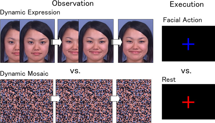
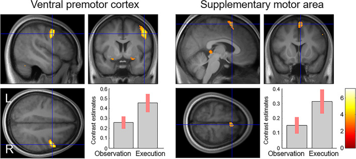
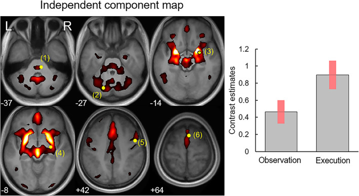
Observing and understanding others' emotional facial expressions, possibly through motor synchronization, plays a primary role in face-to-face communication. To understand the underlying neural mechanisms, previous functional magnetic resonance imaging (fMRI) studies investigated brain regions that are involved in both the observation/execution of emotional facial expressions and found that the neocortical motor regions constituting the action observation/execution matching system or mirror neuron system were active. However, it remains unclear (1) whether other brain regions in the limbic, cerebellum, and brainstem regions could be also involved in the observation/execution matching system for processing facial expressions, and (2) if so, whether these regions could constitute a functional network. To investigate these issues, we performed fMRI while participants observed dynamic facial expressions of anger and happiness and while they executed facial muscle activity associated with angry and happy facial expressions. Conjunction analyses revealed that, in addition to neocortical regions (i.e., the right ventral premotor cortex and right supplementary motor area), bilateral amygdala, right basal ganglia, bilateral cerebellum, and right facial nerve nucleus were activated during both the observation/execution tasks. Group independent component analysis revealed that a functional network component involving the aforementioned regions were activated during both observation/execution tasks. The data suggest that the motor synchronization of emotional facial expressions involves a widespread observation/execution matching network encompassing the neocortex, limbic system, basal ganglia, cerebellum, and brainstem.
Keywords: amygdala; cerebellum; dynamic facial expressions of emotion; facial nerve nucleus; group independent component analysis (ICA); mirror neuron system.
© 2023 The Authors. Human Brain Mapping published by Wiley Periodicals LLC.
Conflict of interest statement
The authors declare no competing financial or other interests.
Figures

Similar articles
-
Positive facial affect - an fMRI study on the involvement of insula and amygdala.PLoS One. 2013 Aug 21;8(8):e69886. doi: 10.1371/journal.pone.0069886. eCollection 2013. PLoS One. 2013. PMID: 23990890 Free PMC article.
-
Widespread and lateralized social brain activity for processing dynamic facial expressions.Hum Brain Mapp. 2019 Sep;40(13):3753-3768. doi: 10.1002/hbm.24629. Epub 2019 May 14. Hum Brain Mapp. 2019. PMID: 31090126 Free PMC article.
-
Affect-specific activation of shared networks for perception and execution of facial expressions.Soc Cogn Affect Neurosci. 2013 Apr;8(4):370-7. doi: 10.1093/scan/nss008. Epub 2012 Jan 24. Soc Cogn Affect Neurosci. 2013. PMID: 22275167 Free PMC article.
-
Functional atlas of emotional faces processing: a voxel-based meta-analysis of 105 functional magnetic resonance imaging studies.J Psychiatry Neurosci. 2009 Nov;34(6):418-32. J Psychiatry Neurosci. 2009. PMID: 19949718 Free PMC article. Review.
-
The shared neural basis of empathy and facial imitation accuracy.Neuroimage. 2014 Jan 1;84:367-75. doi: 10.1016/j.neuroimage.2013.08.061. Epub 2013 Sep 3. Neuroimage. 2014. PMID: 24012546 Review.
Cited by
-
The Impact of the Perception of Primary Facial Emotions on Corticospinal Excitability.Brain Sci. 2023 Sep 6;13(9):1291. doi: 10.3390/brainsci13091291. Brain Sci. 2023. PMID: 37759892 Free PMC article.
-
Cerebellar Contributions to Traumatic Autobiographical Memory in People with Post-Traumatic Stress Disorder.Cerebellum. 2024 Dec;23(6):2332-2340. doi: 10.1007/s12311-024-01731-9. Epub 2024 Aug 24. Cerebellum. 2024. PMID: 39180693
-
Inhibitory control towards angry stimuli in patients with binge eating disorder: a pilot study.J Eat Disord. 2023 Jul 31;11(1):125. doi: 10.1186/s40337-023-00848-2. J Eat Disord. 2023. PMID: 37525245 Free PMC article.
-
Dynamic concordance between subjective and facial EMG hedonic responses during the consumption of gel-type food.Curr Res Food Sci. 2025 Jun 4;10:101107. doi: 10.1016/j.crfs.2025.101107. eCollection 2025. Curr Res Food Sci. 2025. PMID: 40547888 Free PMC article.
-
Sensorimotor regulation of facial expression - An untouched frontier.Neurosci Biobehav Rev. 2024 Jul;162:105684. doi: 10.1016/j.neubiorev.2024.105684. Epub 2024 May 6. Neurosci Biobehav Rev. 2024. PMID: 38710425 Free PMC article. Review.
References
-
- Alahmadi, A. A. S. (2021). Effects of different smoothing on global and regional resting functional connectivity. Neuroradiology, 63, 99–109. - PubMed
-
- Andersson, J. L. , Hutton, C. , Ashburner, J. , Turner, R. , & Friston, K. (2001). Modeling geometric deformations in EPI time series. NeuroImage, 13, 903–919. - PubMed
-
- Ashburner, J. , & Friston, K. J. (2005). Unified segmentation. NeuroImage, 26, 839–851. - PubMed
Publication types
MeSH terms
LinkOut - more resources
Full Text Sources
Miscellaneous
