Combined supracerebellar infratentorial and right occipital interhemispheric approach to falcotentorial junction meningioma: A case report
- PMID: 36895242
- PMCID: PMC9990807
- DOI: 10.25259/SNI_1027_2022
Combined supracerebellar infratentorial and right occipital interhemispheric approach to falcotentorial junction meningioma: A case report
Abstract
Background: Falcotentorial meningioma is a rare tumor of pineal region, arising from the dural folds where the tentorium and falx meet. Due to the deep location and near closeness to significant neurovascular structures, gross-total tumor resection in this area can be complicated. Pineal meningiomas can be resected using a variety of approaches; however, all these approaches are associated with a significant risk of postoperative complications.
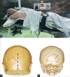
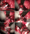
Case description: A 50-year-old female patient who presented with several headaches and visual field defect and diagnosed with pineal region tumor is discussed in the case report. Patient was successfully managed surgically by combined supracerebellar infratentorial and right occipital interhemispheric approach. Cerebrospinal fluid circulation was restored after surgery and neurological defects were regressed.
Conclusion: Our case shows that it is possible to completely remove giant falcotentorial meningiomas with minimal brain retraction, preserve the straight sinus and vein of Galen, and prevent neurological impairments by combining two approaches.
Keywords: Case report; Combined approach; Falcotentorial meningioma; Occipital interhemispheric approach; Pineal region; Supracerebellar infratentorial approach.
Copyright: © 2023 Surgical Neurology International.
Conflict of interest statement
There are no conflicts of interest.
Figures


References
-
- Asari S, Maeshiro T, Tomita S, Kawauchi M, Yabuno N, Kinugasa K, et al. Meningiomas arising from the falcotentorial junction. Clinical features, neuroimaging studies, and surgical treatment. J Neurosurg. 1995;82:726–38. - PubMed
-
- Bassiouni H, Asgari S, König HJ, Stolke D. Meningiomas of the falcotentorial junction: Selection of the surgical approach according to the tumor type. Surg Neurol. 2008;69:339–49. - PubMed
-
- de Andoain GB, Delgado-Fernández J, Cuesta JR, GilSimoes R, Frade-Porto N, Sánchez MP. Meningiomas originated at the falcotentorial region: Analysis of topographic and diagnostic features guiding an optimal surgical planning. World Neurosurg. 2019;123:e723–33. - PubMed
-
- Celtikci E, Nunez M, Liu JK, Gardner PA, Cohen-Gadol AA, Fernandez-Miranda JC. Interhemispheric precuneus retrosplenial transfalcine approach for falcotentorial meningiomas: Anatomic study and clinical series. Operative Neurosurg (Hagerstown) 2021;21:48–56. - PubMed
-
- Ore CL, Magill ST, McDermott MW. Falcotentorial meningiomas. Handb Clin Neurol. 2020;170:107–14. - PubMed
Publication types
LinkOut - more resources
Full Text Sources
