. 2023 Mar;48(2):187-197.
doi: 10.30476/IJMS.2022.93594.2497.

Association of Oxidative Stress with Kidney Injury in a Hyperandrogenemic Female Rat Model

Affiliations

Association of Oxidative Stress with Kidney Injury in a Hyperandrogenemic Female Rat Model

Nima Forghani et al. Iran J Med Sci. 2023 Mar.

Abstract

Background: Polycystic ovary syndrome (PCOS) is the most common reproductive dysfunction in premenopausal women. PCOS is associated with oxidative stress (OS), which is the main risk factor for renal diseases. This study aimed to investigate the mechanisms responsible for renal injury in a hyperandrogenemic female rat model.

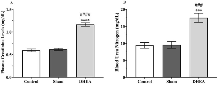
Methods: This study was conducted from December 2019 to September 2021 at Shiraz Nephro-Urology Research Centre, Shiraz University of Medical Sciences (Shiraz, Iran). Thirty female Sprague-Dawley rats were randomly divided into three groups (n=10), namely control, sham, and dehydroepiandrosterone (DHEA). Plasma total testosterone, plasma creatinine (Cr), and blood urea nitrogen (BUN) levels were measured. In addition, total oxidant status (TOS), total antioxidant capacity (TAC), oxidative stress index (OSI), and histopathological changes in the ovaries and kidneys were determined. Data were analyzed using the GraphPad Prism software, and P<0.05 was considered statistically significant.

Results: Plasma total testosterone levels increased by nine-fold in DHEA-treated rats compared to controls (P=0.0001). Administration of DHEA increased Cr and BUN levels and caused severe renal tubular cell injury. In addition, plasma and tissue (kidney and ovary) TAC levels decreased significantly, but TOS levels and OSI values were significantly increased (P=0.019). Significant damage to both glomerular and tubular parts of the kidney and ovarian follicular structure was observed in the DHEA group.

Conclusion: Hyperandrogenemia caused systemic abnormalities through OS-related mechanisms and damaged renal and ovarian tissues. DHEA treatment in rat models is recommended to study the mechanisms that mediate PCOS-associated renal injury.

Keywords: Dehydroepiandrosterone; Kidney disease; Oxidative stress; Polycystic ovary syndrome.


Conflict of interest statement

None declared.

Figures

Figure 1
Figure 1
The figure shows the effect of 28 days of DHEA administration on the level of plasma total testosterone in the control, sham (received 0.2 mL sesame oil subcutaneously per day as a vehicle), and DHEA (subcutaneously injected with 6 mg DHEA per 100 g/day) groups (n=10). The data were analyzed using one-way analysis of variance (ANOVA) with Tukey’s post hoc test and expressed as mean±SEM. **** and #### P<0.0001 represents significant differences between the control and sham groups, respectively. DHEA: Dehydroepiandrosterone.
Figure 2
Figure 2
The figure shows the effects of 28 days of DHEA administration on the level of plasma creatinine (A) and blood urea nitrogen (BUN) (B) in the control, sham (received 0.2 mL sesame oil subcutaneously per day as a vehicle), and DHEA (subcutaneously injected with 6 mg DHEA per 100 g/day) groups (n=10). The data were analyzed using one-way analysis of variance (ANOVA) with Tukey’s post hoc test and expressed as mean±SEM. ***P<0.001 and ****P<0.0001 represent significant differences with the control group, ###P<0.001 and ####P<0.0001 represent significant differences with the sham group. DHEA: Dehydroepiandrosterone.
Figure 3
Figure 3
The figure shows the photomicrograph of ovarian tissue in the experimental groups (H&E staining). (A) The ovarian section of the control group with healthy growing follicles and corpora lutea and (B) a tertiary (Graafian) follicle. (C) The ovarian section of the sham group (received 0.2 mL sesame oil subcutaneously per day as a vehicle) with follicles at the different stages of development and corpora lutea and (D) a tertiary (Graafian) follicle. (E) The ovarian section of the DHEA group (subcutaneously injected with 6 mg DHEA per 100 g/day) with several cyst-like follicles and very few corpora lutea, and (F) cystic follicles. PF: Primary follicle; SF: Secondary follicle; TF: Tertiary (Graafian) follicle; CF: Cystic follicle; CL: Corpus luteum; TT: Theca layer thickness; GT: Granulosa layer thickness; DHEA: Dehydroepiandrosterone; Magnification: ×100 (A, C, E) and ×400 (B, D, F).
Figure 4
Figure 4
The figure shows the effect of 28 days of DHEA administration on (A) tertiary follicle diameter, (B) theca layer thickness, and (C) granulosa layer thickness of the ovary in the control, sham (received 0.2 mL sesame oil subcutaneously per day as a vehicle), and DHEA (subcutaneously injected with 6 mg DHEA per 100 g/day) groups (10 follicles in each section and a total of 20 follicles in each group). The data were analyzed using one-way analysis of variance (ANOVA) with Tukey’s post hoc test and expressed as mean±SEM. *P<0.05 and ****P<0.0001 represent significant differences with the control group. #P<0.05, ##P<0.01 and ####P<0.0001 represent significant differences with the sham group. DHEA: Dehydroepiandrosterone.
Figure 5
Figure 5
The figure shows the photomicrograph of renal tissue in the experimental groups (H&E staining). The renal section of the control group with a completely normal appearance in the (A) cortex and (B) medulla. The renal section of the sham group (received 0.2 mL sesame oil subcutaneously per day as a vehicle) with a normal condition in the (C) cortex and (D) medulla. The renal section of the DHEA group (subcutaneously injected with 6 mg DHEA per 100 g/day) with (E) epithelial cell injury and shedding of brush borders in the convoluted proximal tubules (thin black arrow) and bowman space enlargement (yellow arrowhead) in the cortex and (F) casts inside the tubules (thick black arrow) in the medulla. Magnification: ×400.

Similar articles

Cited by

References

    1. Azziz R, Carmina E, Chen Z, Dunaif A, Laven JS, Legro RS, et al. Polycystic ovary syndrome. Nat Rev Dis Primers. 2016;2:16057. doi: 10.1038/nrdp.2016.57. - DOI - PubMed
    1. Chappell NR, Gibbons WE, Blesson CS. Pathology of hyperandrogenemia in the oocyte of polycystic ovary syndrome. Steroids. 2022;180:108989. doi: 10.1016/j.steroids.2022.108989. [ PMC Free Article ] - DOI - PMC - PubMed
    1. Rotterdam EA-SPcwg. Revised 2003 consensus on diagnostic criteria and long-term health risks related to polycystic ovary syndrome (PCOS) Hum Reprod. 2004;19:41–7. doi: 10.1093/humrep/deh098. - DOI - PubMed
    1. Bruni V, Capozzi A, Lello S. The Role of Genetics, Epigenetics and Lifestyle in Polycystic Ovary Syndrome Development: the State of the Art. Reprod Sci. 2022;29:668–79. doi: 10.1007/s43032-021-00515-4. - DOI - PubMed
    1. Dewailly D, Lujan ME, Carmina E, Cedars MI, Laven J, Norman RJ, et al. Definition and significance of polycystic ovarian morphology: a task force report from the Androgen Excess and Polycystic Ovary Syndrome Society. Hum Reprod Update. 2014;20:334–52. doi: 10.1093/humupd/dmt061. - DOI - PubMed

Publication types

LinkOut - more resources