

Clinical application of VATS combined with 3D-CTBA in anatomical basal segmentectomy
- PMID: 36895493
- PMCID: PMC9989288
- DOI: 10.3389/fonc.2023.1137620
Clinical application of VATS combined with 3D-CTBA in anatomical basal segmentectomy
Abstract
Objective: This study aimed to summarize the clinical application experience of video-assisted thoracic surgery (VATS) combined with three-dimensional computed tomography-bronchography and angiography (3D-CTBA) in anatomical basal segmentectomy.
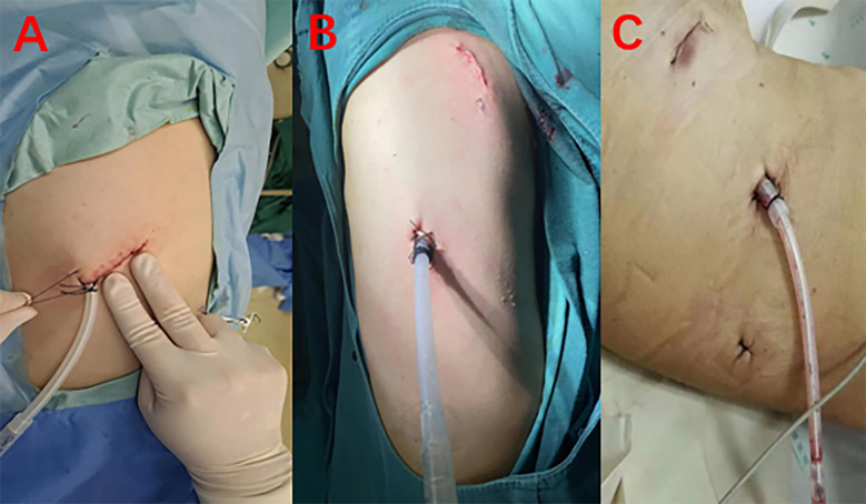
Methods: Clinical data of 42 patients who underwent bilateral lower sub-basal segmentectomy by VATS combined with 3D-CTBA in our hospital from January 2020 to June 2022 were retrospectively analyzed; the patients included 20 males and 22 females, with a median age of 48 (30-65) years. Combined with the preoperative enhanced CT and 3D-CTBA techniques to identify the altered bronchi, arteries, and veins during the operation, the anatomical resection of each basal segment of both lower lungs was completed through the fissure approach or inferior pulmonary vein approach.
Results: All operations were successfully completed without conversion to thoracotomy or lobectomy. The median operation time was 125 (90-176) min, the median intraoperative blood loss was 15 (10-50) mL, the median postoperative thoracic drainage time was 3 (2-17) days, and the median postoperative hospital stay was 5 (3-20) days. The median number of resected lymph nodes was 6 (5-8). There was no in-hospital death. Postoperative pulmonary infection occurred in 1 case, lower extremity deep vein thrombosis (DVT) in 3 cases, pulmonary embolism in 1 case, and persistent air leakage in the chest in 5 cases, all of which were improved by conservative treatment. Two cases of pleural effusion after discharge were improved after ultrasound guided drainage. Postoperative pathology showed 31 cases of minimally invasive adenocarcinoma, 6 cases of adenocarcinoma in situ (AIS), 3 cases of severe atypical adenomatous hyperplasia (AAH), and 2 cases of other benign nodules. All cases were lymph node-negative.
Conclusion: VATS combined with 3D-CTBA is safe and feasible in anatomical basal segmentectomy; consequently, this approach should be promoted and applied in clinical work.
Keywords: 3D-CTBA; VATS; basal segment resection; lung cancer; thoracic surgery.
Copyright © 2023 Zhang, Wang, Feng, Chen, Feng, Qin and Han.
Conflict of interest statement
The authors declare that the research was conducted in the absence of any commercial or financial relationships that could be construed as a potential conflict of interest. The handling editor HL declared a shared parent affiliation with the authors at the time of review.
Figures


References
-
- Hu W, Zhang K, Han X, Zhao J, Wang G, Yuan S, et al. . Three-dimensional computed tomography angiography and bronchography combined with three-dimensional printing for thoracoscopic pulmonary segmentectomy in stage IA non-small cell lung cancer. J Thorac Dis (2021) 13(2):1187–95. doi: 10.21037/jtd-21-16 - DOI - PMC - PubMed
LinkOut - more resources
Full Text Sources