

Use of point-of-care ultrasound to diagnose spontaneous rupture of fibroid in pregnancy
- PMID: 36895497
- PMCID: PMC9979928
- DOI: 10.24908/pocus.v6i1.14757
Use of point-of-care ultrasound to diagnose spontaneous rupture of fibroid in pregnancy
Abstract
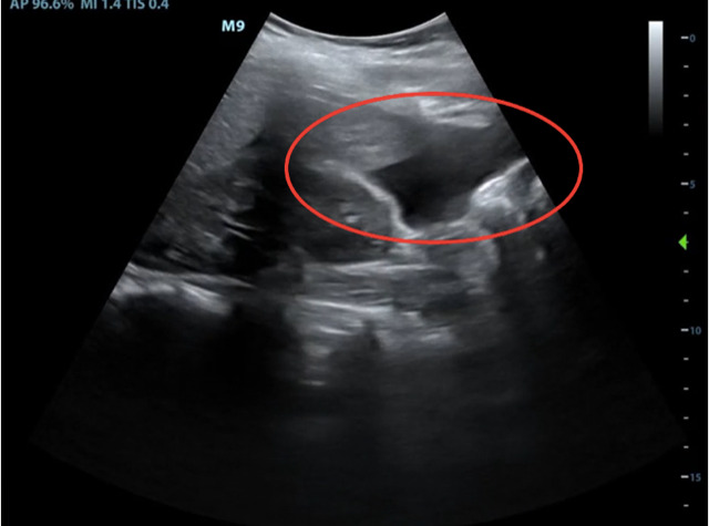
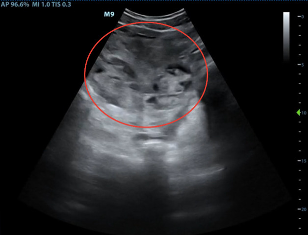
Background: Complications of fibroids in pregnancy are well known, including postpartum hemorrhage, labor dystocia, and cesarean delivery. Outside of pregnancy and labor, the rare occurrence of spontaneous fibroid rupture has been documented. Case: The current case report involves a woman who presented with acute abdominal pain in the third trimester of pregnancy and was found to have spontaneous rupture of a fibroid before the onset of labor. Her initial presentation, diagnosis through use of point-of-care ultrasound, acute surgical management, and postoperative course are described. Conclusion: When assessing acute abdominal pain in a pregnant patient, fibroid rupture should be considered despite the absence of prior uterine surgery. Bedside point-of-care ultrasonography is a useful tool for assessment of abdominal pain in the third trimester of pregnancy.
Keywords: FAST exam; POCUS; fibroid; hemoperitoneum; pregnancy.
Author(s) retain the copyright for their work.
Conflict of interest statement
The authors have no conflicts of interest to declare.
Figures


Similar articles
-
Rare complication of fibroids in pregnancy: Spontaneous fibroid rupture.J Obstet Gynaecol Res. 2017 Sep;43(9):1485-1488. doi: 10.1111/jog.13405. Epub 2017 Jul 10. J Obstet Gynaecol Res. 2017. PMID: 28691348
-
Successful laparoscopic management of acute abdominal pain due to spontaneous rupture of subserosal vessels overlying a uterine fibroid: a case report and surgical video.BMC Womens Health. 2022 Sep 23;22(1):388. doi: 10.1186/s12905-022-01970-0. BMC Womens Health. 2022. PMID: 36138425 Free PMC article.
-
Hemorrhaging Uterine Fibroid Leading to Emergent Early Term Cesarean Delivery: A Case Report.AJP Rep. 2024 Oct 22;14(4):e250-e253. doi: 10.1055/a-2434-5650. eCollection 2024 Jul. AJP Rep. 2024. PMID: 39440246 Free PMC article.
-
A uterine fibroid presenting as an incarcerated epigastric hernia: a case report and review of the literature.J Med Case Rep. 2023 Aug 18;17(1):370. doi: 10.1186/s13256-023-04032-7. J Med Case Rep. 2023. PMID: 37596689 Free PMC article. Review.
-
Delivery for women with a previous cesarean: guidelines for clinical practice from the French College of Gynecologists and Obstetricians (CNGOF).Eur J Obstet Gynecol Reprod Biol. 2013 Sep;170(1):25-32. doi: 10.1016/j.ejogrb.2013.05.015. Epub 2013 Jun 28. Eur J Obstet Gynecol Reprod Biol. 2013. PMID: 23810846 Review.
Cited by
-
A subserosal uterine leiomyoma complicated with intra-abdominal haemorrhage: A case report.Case Rep Womens Health. 2023 Sep 23;39:e00549. doi: 10.1016/j.crwh.2023.e00549. eCollection 2023 Sep. Case Rep Womens Health. 2023. PMID: 37781450 Free PMC article.
-
Intra-Abdominal Hemorrhagic Catastrophe due to Large Subserous Myomatous Capsular Venous Rupture.Case Rep Obstet Gynecol. 2022 Jan 4;2022:2696213. doi: 10.1155/2022/2696213. eCollection 2022. Case Rep Obstet Gynecol. 2022. PMID: 35036016 Free PMC article.
References
Publication types
LinkOut - more resources
Full Text Sources