Case Reports
. 2023 Feb 4;15(2):e34636.
doi: 10.7759/cureus.34636. eCollection 2023 Feb.

Ten-Fraction Stereotactic Radiosurgery With Different Gross Tumor Doses and Inhomogeneities for Brain Metastasis of >10 cc: Treatment Responses Suggesting Suitable Biological Effective Dose Formula for Single and 10 Fractions

Affiliations
Case Reports

Ten-Fraction Stereotactic Radiosurgery With Different Gross Tumor Doses and Inhomogeneities for Brain Metastasis of >10 cc: Treatment Responses Suggesting Suitable Biological Effective Dose Formula for Single and 10 Fractions

Kazuhiro Ohtakara et al. Cureus. .

Abstract

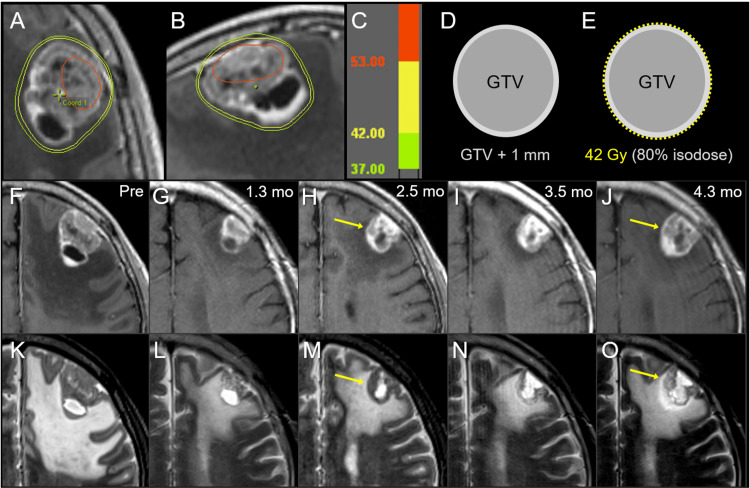
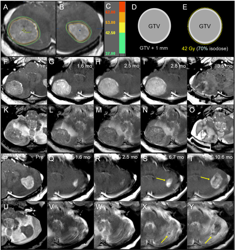
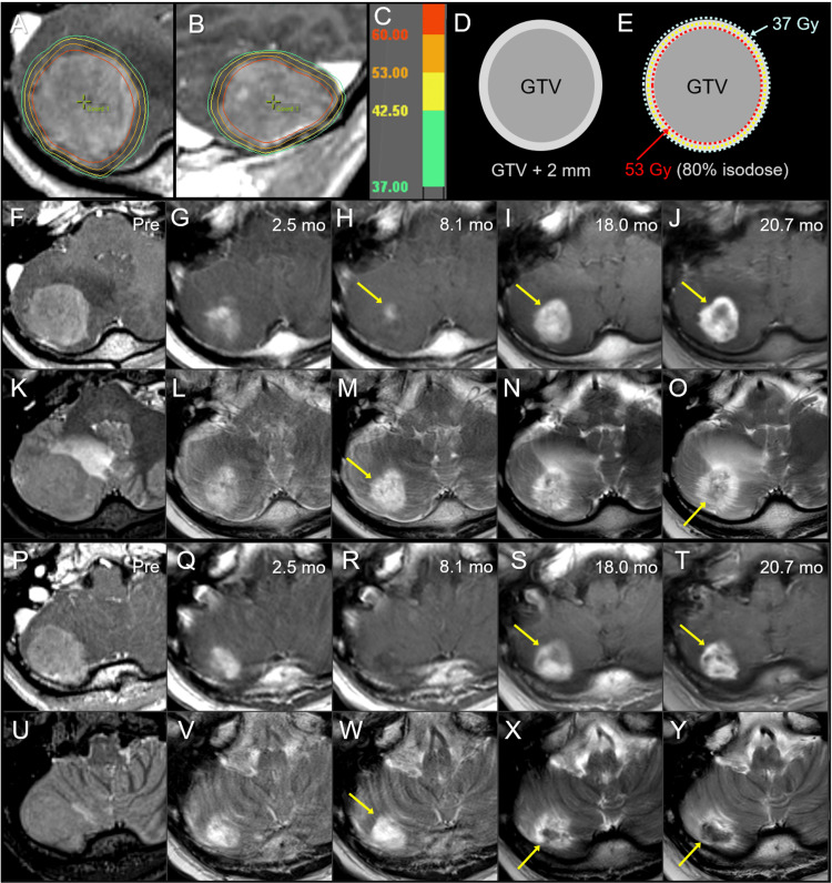
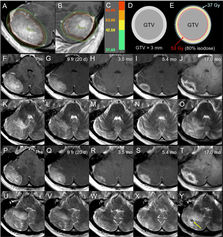
Stereotactic radiosurgery (SRS) with >5 fractions (fr) has been increasingly adopted to improve local control and safety for brain metastases (BM) of >10 cm3, given the limited brain tolerance of SRS with ≤5 fr. However, the optimal indication and treatment design, including the prescribed dose and distribution for 10 fr SRS, remains uncertain. A single fr of 24 Gy provides approximately 95% of the one-year local tumor control probability. The potential SRS doses in 10 fr that is clinically equivalent to a single fr of 24 Gy regarding anti-tumor effect range from 48.4 to 81.6 Gy as biological effective doses (BED) as a function of the BED model formulas along with the alpha/beta ratios. The most appropriate BED formula in conjunction with an alpha/beta ratio to estimate similar anti-BM effects for single and 10 fr remains controversial. Herein, we describe four cases of symptomatic radiation-naïve BM >10 cm3 (range, 11 to 26 cm3), treated with 10 fr SRS with a standard prescribed dose of 42 Gy, for which modified dynamic conformal arcs were used with forward planning to improve dose conformity. In the first two cases with gross tumor volumes (GTV) of 15.3 and 10.9 cm3, 42 Gy was prescribed to 70%-80% isodose, normalized to 100% at the isocenter, which encompasses the boundary of the planning target volume: GTV + isotropic 1 mm margin. The tumor responses were initially marked regression followed by regrowth within three months in case 1 and no shrinkage with subsequent progression within three months in case 2. In the remaining two cases with larger GTVs of 19.1 and 26.2 cm3, the GTV boundary and 2-3 mm margin-added object volume was covered by 80% and 56% isodoses with 53 Gy and 37 Gy, respectively, to further increase the marginal and internal doses of GTV and to ensure moderate dose spillage outside the GTV, while >1-1.5 mm outside the GTV was covered by 42 Gy with 63% isodose. According to the BED based on the linear-quadratic (LQ) model with an alpha/beta ratio of 10 (BED10), 53 Gy corresponds to approximately 81 Gy in BED10 and 24 Gy in a single fr. Excellent initial maximum tumor response and subsequently sustained tumor regression (STR) were achieved in both cases. Subsequently, enlarging nodules that could not exclude the possibility of tumor regrowth were disclosed within two years, while late adverse radiation effects remained moderate. These dose-effect relationships suggest that a GTV marginal dose of ≥53 Gy with ≤80% isodose would be preferred to effect ≥1-year STR and that further dose escalation of both marginal and internal GTV may be necessary to achieve ≥2-year STR, while GTV of >25 cm3 may be unsuitable for 10 fr SRS in terms of long-term brain tolerance. Among LQ, LQ-cubic, and LQ-linear model formulas and alpha/beta ratios of 10-20, BED10 may be clinically most suitable to estimate a 10 fr SRS dose that provides anti-BM efficacy similar to that for a single fr.

Keywords: alpha/beta ratio; biological effective dose; brain metastasis; dynamic conformal arcs; fractionation; large tumor; linear-quadratic model; linear-quadratic-cubic model; linear-quadratic-linear model; stereotactic radiosurgery.


Conflict of interest statement

The authors have declared that no competing interests exist.

Figures

Figure 1
Figure 1. Forward planning method for modified dynamic conformal arcs.
The images show target volume definition consisting of gross tumor volume (GTV) and planning target volume (PTV) (A); PTV covering by prescribed isodose surface (IDS) in general dynamic conformal arcs (DCA) (B); modified GTV (mGTV) compared to the original GTV (C); and PTV covering by prescribed IDS in modified DCA (mDCA) (D). (B) In this case, when the original GTV is directly used as an object volume for leaf adaptation, the dose conformity of the PTV with the prescribed IDS (dashed line) has room for improvement, that is, over-coverage. (C) To improve the dose conformity, a copy of the original GTV is used to generate a surrogate object for leaf adaptation to be deformed in a desired shape, namely, the mGTV, and then modified according to the over- and/or under-coverage of the PTV with the initial prescribed IDS. (D) When an appropriately modified mGTV is used for leaf adaptation, the dose conformity of the PTV with the prescribed IDS is improved.
Figure 2
Figure 2. Case 1: Dose distribution, planning design, and images before and after 10-fraction stereotactic radiosurgery.
The images show (A, B) dose distributions (A: axial view; B: sagittal view); corresponding isodoses (C); schema of the target definition, including GTV and PTV: GTV + isotropic 1 mm (D); schema of treatment planning (E); contrast-enhanced (CE) T1-weighted images (T1-WI) (F-J); and T2-weighted images (T2-WI) (K-O); 4 days before the initiation of stereotactic radiosurgery (SRS) (Pre) (F, K); at 1.3 months (mo) after the initiation of SRS (G, L); at 2.5 months (H, M); at 3.5 months (I, N); and 4.3 months (J, O). (A) The irradiated tumor volume of 53 Gy (red) is extremely limited. (E) A dose of 42 Gy is prescribed for 80% IDS, normalized to 100% at the isocenter, encompassing the PTV boundary. (F-O) On T1/T2 matching, tumor regrowth is observed once at 2.5 months (arrows in H, M) and again at 4.3 months (arrows in J, O), while tumor regression is markedly observed at 1.3 months and slightly at 3.5 months. GTV: gross tumor volume; PTV: planning target volume; IDS: isodose surface
Figure 3
Figure 3. Case 2: Dose distribution, planning design, and images before and after 10-fraction stereotactic radiosurgery.
The images show (A, B) axial views of dose distributions in the right (A) and left (B) cerebellar lesions; corresponding isodoses (C); schema of the GTV and PTV: GTV + 1 mm (D); schema of treatment planning (E); the right cerebellar lesion (F-O); the left cerebellar lesion (P-Y); CE-T1-WI (F-J, P-T); T2-WI (K-O, U-Y); six days before the initiation of SRS (Pre) (F, K, P, U); at 1.6 months (mo) after the initiation of SRS (G, L, Q, V); at 2.5 months (H, M, R, W); at 2.8 months (I, N); at 3.5 months (J, O); at 6.7 months (S, X); and 10.6 months (T, Y). (A, B) The substantial volumes of both GTVs are covered by 53 Gy (orange). (E) 42 Gy is prescribed to 70% IDS covering the PTV. (F-O) The right cerebellar lesion showed no regression at 1.6 months (G, L), with subsequent enlargement of up to 39 mm in maximum diameter at 2.8 months (I, N). (P-Y) The left cerebellar lesion achieved a nearly complete remission at 2.5 months (Q, R, V, W); however, enlargement of both the enhancing lesion and iso-intensity to a low-intensity nodule on T2-WI are observed at 6.7-10.6 months (S, T, X, Y). GTV: gross tumor volume; PTV: planning target volume; CE: contrast-enhanced; T1-WI: T1-weighted image; T2-WI: T2-weighted image; SRS: stereotactic radiosurgery; IDS: isodose surface
Figure 4
Figure 4. Case 3: Dose distribution, planning design, and images before and after 10-fraction stereotactic radiosurgery.
The images show (A, B) dose distributions (A: axial view; B: sagittal view); corresponding isodoses (C); schema of GTV and object volume for dose evaluation: GTV + 2 mm (D); schema of treatment planning (E); axial views at the plane of initial GTV center (F-O); axial views at the more caudal plane (P-Y); CE-T1-WI (F-J, P-T); T2-WI (K-O, U-Y); six days before the initiation of SRS (Pre) (F, K, P, U); at 2.5 months (mo) after the initiation of SRS (G, L, Q, V); at 8.1 months (H, M, R, W); at 18.0 months (I, N, S, X); and at 20.7 months (J, O, T, Y). (A, B) The D66% of the GTV is 62 Gy (red), in which DX% indicates the dose irradiated to at least X% of the volume. (E) 53 Gy is prescribed to 80% IDS encompassing the GTV boundary, and the GTV + 2 mm boundary is simultaneously covered by 37 Gy. (G, L, Q, V) At 2.5 months, marked tumor regression is observed in which the iso-intensity mass in T2-WI almost disappeared, leaving a slight enhancement. (H, M, R, W) Subsequently, a marked discrepancy, i.e., T1/T2 mismatch, between the slightly enhancing lesion (arrow in H) and high-intensity tumor necrosis (arrows in M, W) is observed at 8.1 months. (I, N, S, X) At 18.0 months, enlargement of the enhancing lesion (arrows in I, S) up to the high-intensity lesion boundary on T2-WI and a low-intensity nodule on T2-WI (arrow in X) appeared. (J, O, T, Y) At 20.7 months, the low-intensity nodule (arrow in Y) enlarged, and an iso-intensity irregular mass (arrow in O) became noticeable, while the enhancing lesion (arrows in I, S), suggestive of the radiation effect, did not show significant enlargement (arrows in J, T). GTV: gross tumor volume; CE: contrast-enhanced; T1-WI: T1-weighted image; T2-WI: T2-weighted image; SRS: stereotactic radiosurgery; IDS: isodose surface
Figure 5
Figure 5. Case 4: Dose distribution, planning design, and images before and after 10-fraction stereotactic radiosurgery.
The images show (A, B) dose distributions (A: axial view; B: sagittal view); corresponding isodoses (C); schema of GTV and object volume for dose evaluation: GTV + 3 mm (D); schema of treatment planning (E); axial views at the plane of initial GTV center (F-O); axial views at the more caudal plane (P-Y); CE-T1-WI (F-J, P-T); T2-WI (K-O, U-Y); 10 days before the initiation of SRS (Pre) (F, K, P, U); at nine fractions (fr) and also 20 days (d) after pre-image acquisition (G, L, Q, V); at 3.5 months (mo) after the initiation of SRS (H, M, R, W); at 5.4 months (I, N, S, X); and at 17.0 months (J, O, T, Y). (A, B) The D85% of GTV is 62 Gy (red); (E) 53 Gy is prescribed for 80% IDS that fully encompasses the GTV boundary, and the GTV + 3 mm boundary is simultaneously covered by 37 Gy, given the presumed brain invasion and the 10-day interval between image acquisition and SRS initiation. (G, L, Q, V) Slight tumor regression is observed at 9 fr. (G, L, Q, V) A marked decrease in the enhancing lesion and a visible mass on T2-WI are observed at 3.5 months. (I, N, S, X) The increase in enhancement without enlarging the nodule on T2-WI suggested a radiation effect at 5.4 months. (J, O, T, Y) Further enlargement of the enhancing lesion, a low-intensity nodule on T2-WI (arrow in Y), and aggravation of perilesional edema are observed at 17.0 months. GTV: gross tumor volume; CE: contrast-enhanced; T1-WI: T1-weighted image; T2-WI: T2-weighted image; SRS: stereotactic radiosurgery; IDS: isodose surface

References

    1. Surgical resection versus stereotactic radiosurgery on local recurrence and survival for patients with a single brain metastasis: a systematic review and meta-analysis. González L, Castro S, Villa E, Zomosa G. Br J Neurosurg. 2021;35:703–713. - PubMed
    1. Update on the management of brain metastasis. Singh K, Saxena S, Khosla AA, McDermott MW, Kotecha RR, Ahluwalia MS. Neurotherapeutics. 2022;19:1772–1781. - PMC - PubMed
    1. Tumor control probability of radiosurgery and fractionated stereotactic radiosurgery for brain metastases. Redmond KJ, Gui C, Benedict S, et al. Int J Radiat Oncol Biol Phys. 2021;110:53–67. - PubMed
    1. Single- and multifraction stereotactic radiosurgery dose/volume tolerances of the brain. Milano MT, Grimm J, Niemierko A, et al. Int J Radiat Oncol Biol Phys. 2021;110:68–86. - PMC - PubMed
    1. Significance of target location relative to the depth from the brain surface and high-dose irradiated volume in the development of brain radionecrosis after micromultileaf collimator-based stereotactic radiosurgery for brain metastases. Ohtakara K, Hayashi S, Nakayama N, Ohe N, Yano H, Iwama T, Hoshi H. J Neurooncol. 2012;108:201–209. - PubMed

Publication types

LinkOut - more resources