Review
. 2023 Feb 21:14:1085540.
doi: 10.3389/fneur.2023.1085540. eCollection 2023.

An overview of clinical cerebral microdialysis in acute brain injury

Affiliations
Review

An overview of clinical cerebral microdialysis in acute brain injury

Matthew G Stovell et al. Front Neurol. .

Abstract

Cerebral microdialysis may be used in patients with severe brain injury to monitor their cerebral physiology. In this article we provide a concise synopsis with illustrations and original images of catheter types, their structure, and how they function. Where and how catheters are inserted, their identification on imaging modalities (CT and MRI), together with the roles of glucose, lactate/pyruvate ratio, glutamate, glycerol and urea are summarized in acute brain injury. The research applications of microdialysis including pharmacokinetic studies, retromicrodialysis, and its use as a biomarker for efficacy of potential therapies are outlined. Finally, we explore limitations and pitfalls of the technique, as well as potential improvements and future work that is needed to progress and expand the use of this technology.

Keywords: brain injury; cerebral microdialysis; cerebral physiology; neurocritical care; subarachnoid hemorrhage (SAH); traumatic brain injury (TBI).


Conflict of interest statement

PH was a director of Technicam (Newton Abbot, UK), the manufacturer of the cranial access device used in Cambridge. The remaining authors declare that the research was conducted in the absence of any commercial or financial relationships that could be construed as a potential conflict of interest.

Figures

Figure 1
Figure 1
Cerebral microdialysis from macroscopic to microscopic. (A) Multimodality monitoring in neurocritical care; Illustration of neurocritical care multimodality monitoring including intracranial pressure (ICP), brain tissue oxygen (PbtO2) and microdialysis with an ISCUS/ISCUSflex which are employed alongside standard intensive care monitoring of systemic physiology. Illustration copyright Susan Giorgi-Coll and reproduced here with her permission. (B) Schematic of cerebral microdialysis; 100 kDa cut-off microdialysis catheter inserted into patient's brain, with magnified view of catheter tip demonstrating semipermeable membrane. A gold filament (0.13 x 3 mm) along the end of the catheter allows its identification on CT scans and some MRI sequences. The microdialysis perfusate is pumped slowly (0.3 μL/min) along the outer lumen, the final 10 mm of which is a semipermeable membrane (100 kDa cut-off, outer pore diameter ≈ 2–5 μm). Small molecules freely exchange with the brain interstitium, approaching equilibrium by the time they reach the recovery hole in the end of the central, non-porous channel, where the fluid passes back for collection in microdialysis vials. Glucose, lactate, pyruvate, glycerol, glutamate and urea may be used for clinical monitoring, illustrated to the left of the expanded window. Catheters can also be used to “dose” the surrounding brain with research substrates (including those with stable isotope 13C labeling) and recover the resultant products, illustrated to the right of the expanded window. (C) Scanning electron micrograph of M Dialysis 71 microdialysis catheter sectioned transversely; Upper panel the catheter semipermeable membrane (polyarylethersulfone) external surface demonstrating pores several micrometers across. (B) The central polyurethane non-porous inner channel removed from inside a catheter. Originally published in Helmy et al. (2). Copyright Mary Ann Liebert, Inc. and reproduced with permission (2). (D) Schematic of stylized cell with (enlarged) central mitochondrion illustrating principal metabolites and pathways of energy metabolism examined by cerebral microdialysis. Glucose, pyruvate, and lactate are important species in glycolysis and the TCA cycle for energy metabolism and the regeneration of ATP in conjunction with oxidative phosphorylation and the electron transport chain. Glutamate is the principle excitatory neurotransmitter in the brain and glycerol is a key component of cell membranes. All can be interrogated with clinical microdialysis (highlighted yellow). Glycolysis, the pentose phosphate pathway, the TCA cycle and succinate and glutamine metabolism have been investigated in research studies using microdialysis and retromicrodialysis (highlighted light green). GLUT, glucose transporter; NAD+/NADH, nicotinamide adenine dinucleotide; ADP, adenosine diphosphate; ATP, adenosine triphosphate; Pi, inorganic phosphorous; PCr, phosphocreatine; Cr, creatine; NADP+/NADPH, nicotinamide adenine dinucleotide phosphate; LDH, lactate dehydrogenase; TCA, tricarboxylic acid.
Figure 2
Figure 2
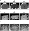
Illustrative examples of multimodal monitoring appearance on CT and MRI. Reformatted coronal slices demonstrate ICP, PbtO2 and microdialysis probes in a patient's brain. (A) CT; The ICP probe's thin shaft and bulbous end can be distinguished from the PbtO2 probe's uniform shaft and tip. Only the gold filament at the end of the microdialysis catheter is visible. (B) MRI T2-weighted SWI sequence; The same bulbous tip of the ICP probe and uniform thickness PbtO2 probe is revealed as with CT. The gold tipped microdialysis catheter is just visible on SWI sequences (highlighted by white dashed circle). (C) MRI T1-weighted MP-RAGE sequence; The same bulbous tip of the ICP probe and uniform thickness PbtO2 probe is revealed as with CT and SWI. The gold tipped microdialysis catheter is not visible. CT, computed tomography; SWI, susceptibility weighted imaging; MP-RAGE, T1-weighted magnetization-prepared rapid gradient-echo; ICP, intracranial pressure; PbtO2, brain tissue oxygen tension; MD, microdialysis.

References

    1. Hutchinson PJ, Jalloh I, Helmy A, Carpenter KLH, Rostami E, Bellander BM, et al. . Consensus statement from the 2014 international microdialysis forum. Intensive Care Med. (2015) 41:1517–28. 10.1007/s00134-015-3930-y - DOI - PMC - PubMed
    1. Helmy A, Carpenter KLH, Skepper JN, Kirkpatrick PJ, Pickard JD, Hutchinson PJ. Microdialysis of cytokines methodological considerations, scanning electron microscopy, and determination of relative recovery. J Neurotrauma. (2009) 26:549–61. 10.1089/neu.2008.0719 - DOI - PubMed
    1. Helmy A, Carpenter KL, Hutchinson PJ. Microdialysis in the human brain and its potential role in the development and clinical assessment of drugs. Curr Med Chem. (2007) 14:1525–37. 10.2174/092986707780831113 - DOI - PubMed
    1. Hillman J, Aneman O, Anderson C, Sjögren F, Säberg C, Mellergård P, et al. . microdialysis technique for routine measurement of macromolecules in the injured human brain. Neurosurgery. (2005) 56:1264–8. 10.1227/01.NEU.0000159711.93592.8D - DOI - PubMed
    1. Giorgi-Coll S, Thelin EP, Lindblad C, Tajsic T, Carpenter KLH, Hutchinson PJA, et al. . Dextran 500 improves recovery of inflammatory markers: an in vitro microdialysis study. J Neurotrauma. (2020) 37:106–14. 10.1089/neu.2019.6513 - DOI - PMC - PubMed

LinkOut - more resources