Recovery of Severe Acute Kidney Injury in a Patient with COVID-19: Role of Lung Ultrasonography
- PMID: 36896101
- PMCID: PMC9994289
- DOI: 10.24908/pocus.v7iKidney.15343
Recovery of Severe Acute Kidney Injury in a Patient with COVID-19: Role of Lung Ultrasonography
Abstract
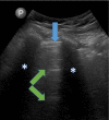
Acute kidney injury (AKI) is recognized as a complication of COVID-19 among hospitalized patients. Lung ultrasonography (LUS) can be a useful tool in the management of COVID-19 pneumonia when interpreted correctly. However, the role of LUS in management of severe AKI in the setting of COVID-19 remains to be defined. We report a 61-year-old male who was hospitalized with acute respiratory failure from COVID-19 pneumonia. In addition to requiring invasive mechanical ventilation, our patient developed AKI and severe hyperkalemia requiring urgent dialytic therapy during his hospital stay. Our patient remained dialysis dependent despite subsequent recovery of lung function. Three days following discontinuation of mechanical ventilation, our patient developed a hypotensive episode during his maintenance hemodialysis treatment. A point of care LUS performed soon after the intradialytic hypotensive episode found no extravascular lung water. Hemodialysis was discontinued and the patient was initiated on intravenous fluids for one week. AKI subsequently resolved. We consider LUS an important tool in identifying COVID-19 patients that would benefit from intravenous fluids following recovery of lung function.
Keywords: AKI; COVID-19; lung ultrasound; point of care ultrasound.
Copyright (c) 2022 Varun Madireddy, Daniel W. Ross, Deepa A. Malieckal, Shamir Hasan, Azzour Hazzan, Hitesh H. Shah.
Conflict of interest statement
The authors have no conflicts of interest or funding to report.
Figures
References
-
- Syndrome Network, Acute Respiratory Distress, Wheeler A P, Bernard G R, Thompson B T, Hayden D, Deboisblanc B, et al. Comparison of two fluid-management strategies in acute lung injury. N Engl j Med. 2006;354(24):2564–2575. - PubMed
-
- Agricola E, Bove T, Oppizzi M, Marino G, Zangrillo A, Margonato A, Picano E. Ultrasound comet-tail images”: a marker of pulmonary edema: a comparative study with wedge pressure and extravascular lung water. Chest. 2005;127(5):1690–1695. - PubMed
Publication types
LinkOut - more resources
Full Text Sources

