

POCUS in Intensive Care Nephrology
- PMID: 36896116
- PMCID: PMC9994305
- DOI: 10.24908/pocus.v7iKidney.15016
POCUS in Intensive Care Nephrology
Abstract
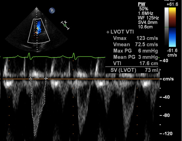
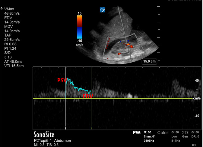
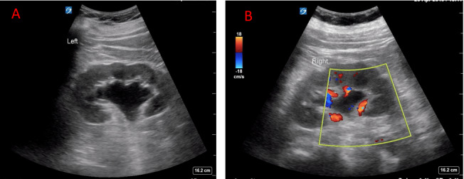
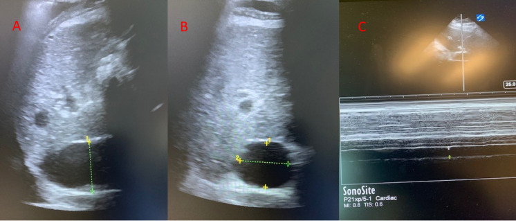
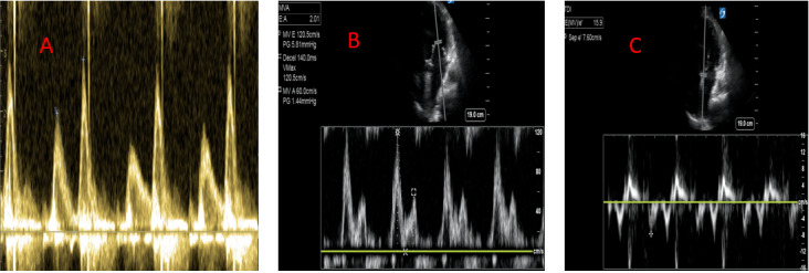
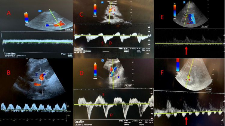
Acute kidney injury (AKI) is a significant problem for patients admitted to the intensive care unit (ICU), both due to the high incidence and associated mortality with rates of AKI requiring renal replacement therapy (RRT) of over 5%, and mortality rates with AKI of over 60% 1, 2.Ultrasound can be used to identify those at risk for AKI and assist with AKI management. Risk factors for AKI in the ICU not only include hypoperfusion but also venous congestion and volume overload. Volume overload and vascular congestion are associated with multi-organ dysfunction and worse renal outcomes. Daily and overall fluid balance, daily weights, and physical examination for edema can be inaccurate and belie true systemic venous pressure 3, 4, 5. Bedside ultrasound allows providers to evaluate vascular flow patterns and obtain a more reliable evaluation of volume status to guide and individualize therapies. Cardiac, lung, and vascular patterns on ultrasound can identify preload responsiveness, which should be assessed to safely manage ongoing fluid resuscitation and assess for signs of fluid intolerance. Here we present an overview in the use of point of care ultrasound with particular emphasis on nephro-centric strategies, namely in the identification of the type of renal injury, renal vascular flow assessment, the static measure of volume status, as well as dynamic evaluation for volume optimization in critically ill patients.
Keywords: AKI; ICU; Intensive care; Nephrology; POCUS; Ultrasound; acute kidney injury.
Copyright (c) 2022 Randi Connor-Schuler, Jonathan Suarez.
Conflict of interest statement
The authors have no conflicts of interest to declare.
Figures
Similar articles
-
Point-of-Care Ultrasound in Nephrology: Beyond Kidney Ultrasound.Diagnostics (Basel). 2025 Jan 27;15(3):297. doi: 10.3390/diagnostics15030297. Diagnostics (Basel). 2025. PMID: 39941227 Free PMC article. Review.
-
Cardiac biomarkers are associated with maximum stage of acute kidney injury in critically ill patients: a prospective analysis.Crit Care. 2017 Apr 12;21(1):88. doi: 10.1186/s13054-017-1674-5. Crit Care. 2017. PMID: 28399905 Free PMC article.
-
RIFLE-based data collection/management system applied to a prospective cohort multicenter Italian study on the epidemiology of acute kidney injury in the intensive care unit.Blood Purif. 2011;31(1-3):159-71. doi: 10.1159/000322161. Epub 2011 Jan 10. Blood Purif. 2011. PMID: 21228585
-
Quantifying systemic congestion with Point-Of-Care ultrasound: development of the venous excess ultrasound grading system.Ultrasound J. 2020 Apr 9;12(1):16. doi: 10.1186/s13089-020-00163-w. Ultrasound J. 2020. PMID: 32270297 Free PMC article.
-
Acute kidney injury in burn patients admitted to the intensive care unit: a systematic review and meta-analysis.Crit Care. 2020 Jan 2;24(1):2. doi: 10.1186/s13054-019-2710-4. Crit Care. 2020. PMID: 31898523 Free PMC article.
Cited by
-
The use of ultrasonography in the diagnosis and management of Acute Kidney Injury and other forms renal dysfunction in the paediatric population: Systematic review protocol.PLoS One. 2025 Jun 18;20(6):e0325549. doi: 10.1371/journal.pone.0325549. eCollection 2025. PLoS One. 2025. PMID: 40531948 Free PMC article.
-
Anatomic evaluation of the height of the carotid bifurcation by 4th year medical students using vascular ultrasonography.J Vasc Bras. 2025 Apr 18;24:e20240111. doi: 10.1590/1677-5449.202401112. eCollection 2025. J Vasc Bras. 2025. PMID: 40275945 Free PMC article.
References
-
- Uchino S, Kellum J A, Bellomo R, Doig G S, Morimatsu H, Morgera S, Schetz M, Tan I, Bouman C, Macedo E, Gibney N, Tolwani A, Ronco C. Beginning and Ending Supportive Therapy for the Kidney (BEST Kidney) Investigators. Acute renal failure in critically ill patients: a multinational, multicenter study. JAMA. 2005;294:813–818. - PubMed
-
- Perren A, Markmann M, Merlani G, Marone C, Merlani P. Fluid balance in critically ill patients. Should we really rely on it? Minerva Anesthesiol. 2011;77:802–811. - PubMed
-
- Flentje K M, Knight C L, Stromfeldt I, Chakrabarti A, Friedman N D. Recording patient bodyweight in hospitals: are we doing well enough? Intern Med J. 2018;48:124–128. - PubMed
-
- Cook D J, Simel D L. The rational clinical examination. Does this patient have abnormal central venous pressure? JAMA. 1996;275:630–634. - PubMed
Publication types
LinkOut - more resources
Full Text Sources