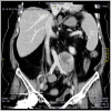
Radio-pathological diagnosis of a retroperitoneal cavernous hemangioma
- PMID: 36896163
- PMCID: PMC9991573
- DOI: 10.1093/jscr/rjad095
Radio-pathological diagnosis of a retroperitoneal cavernous hemangioma
Abstract
Retroperitoneal cavernous hemangioma (RCH) is a rare benign vascular malformation. Only a few cases of RCH were reported. Here we present a case of RCH in a 66-year-old female complaining of long-standing progressive dull abdominal pain.
Keywords: Abdominal mass; Hemangioma; Retroperitoneal Space.
Published by Oxford University Press and JSCR Publishing Ltd. © The Author(s) 2023.
Figures


Similar articles
-
Retroperitoneal cavernous hemangioma misdiagnosed as lymphatic cyst: A case report and review of the literature.World J Clin Cases. 2023 May 26;11(15):3560-3570. doi: 10.12998/wjcc.v11.i15.3560. World J Clin Cases. 2023. PMID: 37383918 Free PMC article.
-
Primary retroperitoneal cavernous hemangioma: A case report and review of the literature.Urol Case Rep. 2024 Feb 26;54:102691. doi: 10.1016/j.eucr.2024.102691. eCollection 2024 May. Urol Case Rep. 2024. PMID: 38516175 Free PMC article.
-
[A Case of Retroperitoneal Cavernous Hemangioma Difficult to Differentiate from Retroperitoneal Liposarcoma].Hinyokika Kiyo. 2017 Dec;63(12):521-524. doi: 10.14989/ActaUrolJap_63_12_521. Hinyokika Kiyo. 2017. PMID: 29370663 Japanese.
-
Imaging findings of retroperitoneal anastomosing hemangioma: a case report and literature review.BMC Urol. 2022 May 22;22(1):77. doi: 10.1186/s12894-022-01022-7. BMC Urol. 2022. PMID: 35599311 Free PMC article. Review.
-
[Retroperitoneal cavernous hemangioma: a case report].Hinyokika Kiyo. 1991 Jul;37(7):725-8. Hinyokika Kiyo. 1991. PMID: 1927773 Review. Japanese.
References
-
- Weidenfeld J, Zakai BB, Faermann R, Barshack I, Aviel-Ronen S. Hemangioma of pancreas: a rare tumor of adulthood. Isr Med Assoc J 2011;13:512–4. - PubMed
-
- England RJ, Woodley H, Cullinane C, McClean P, Walker J, Stringer MD. Pediatric pancreatic hemangioma: a case report and literature review. JOP 2006;7:496–501. - PubMed
-
- Ojili V, Tirumani SH, Gunabushanam G, Nagar A, Surabhi VR, Chintapalli KN, et al. Abdominal hemangiomas: a pictorial review of unusual, atypical, and rare types. Can Assoc Radiol J 2013;64:18–27. - PubMed
Publication types
LinkOut - more resources
Full Text Sources

