. 2023 Feb 21:14:1077798.
doi: 10.3389/fendo.2023.1077798. eCollection 2023.

Serotonin and the ventilatory effects of etonogestrel, a gonane progestin, in a murine model of congenital central hypoventilation syndrome

Affiliations

Serotonin and the ventilatory effects of etonogestrel, a gonane progestin, in a murine model of congenital central hypoventilation syndrome

Alexis Casciato et al. Front Endocrinol (Lausanne). .

Abstract

Introduction: Congenital Central Hypoventilation Syndrome, a rare disease caused by PHOX2B mutation, is associated with absent or blunted CO2/H+ chemosensitivity due to the dysfunction of PHOX2B neurons of the retrotrapezoid nucleus. No pharmacological treatment is available. Clinical observations have reported non-systematic CO2/H+ chemosensitivity recovery under desogestrel.

Methods: Here, we used a preclinical model of Congenital Central Hypoventilation Syndrome, the retrotrapezoid nucleus conditional Phox2b mutant mouse, to investigate whether etonogestrel, the active metabolite of desogestrel, led to a restoration of chemosensitivity by acting on serotonin neurons known to be sensitive to etonogestrel, or retrotrapezoid nucleus PHOX2B residual cells that persist despite the mutation. The influence of etonogestrel on respiratory variables under hypercapnia was investigated using whole-body plethysmographic recording. The effect of etonogestrel, alone or combined with serotonin drugs, on the respiratory rhythm of medullary-spinal cord preparations from Phox2b mutants and wildtype mice was analyzed under metabolic acidosis. c-FOS, serotonin and PHOX2B were immunodetected. Serotonin metabolic pathways were characterized in the medulla oblongata by ultra-high-performance liquid chromatography.

Results: We observed etonogestrel restored chemosensitivity in Phox2b mutants in a non-systematic way. Histological differences between Phox2b mutants with restored chemosensitivity and Phox2b mutant without restored chemosensitivity indicated greater activation of serotonin neurons of the raphe obscurus nucleus but no effect on retrotrapezoid nucleus PHOX2B residual cells. Finally, the increase in serotonergic signaling by the fluoxetine application modulated the respiratory effect of etonogestrel differently between Phox2b mutant mice and their WT littermates or WT OF1 mice, a result which parallels with differences in the functional state of serotonergic metabolic pathways between these different mice.

Discussion: Our work thus highlights that serotonin systems were critically important for the occurrence of an etonogestrel-restoration, an element to consider in potential therapeutic intervention in Congenital Central Hypoventilation Syndrome patients.

Keywords: CO2/H+ chemosensitivity; central breathing disorder; mouse model; progestin; serotoninergic systems.


Conflict of interest statement

The authors declare that the research was conducted in the absence of any commercial or financial relationships that could be construed as a potential conflict of interest.

Figures

Figure 1
Figure 1
Schematic representation of the pharmacological protocols used. Flowchart of the protocols used in ex vivo medulla-spinal cord preparations from Phox2b mutants, wildtype littermates and wildtype Swiss OF1 mice (A). Briefly, after a period allowing fR to stabilize, preparations were subjected to one of four protocols: 1) preparations were exposed to drug-free normal-pH and metabolic acidosis conditions, 2) preparations were exposed to drug-free normal-pH followed by exposure to etonogestrel under normal-pH and then metabolic acidosis conditions, 3) preparations were exposed to drug-free normal-pH followed by exposure to fluoxetine under normal-pH and then metabolic acidosis conditions, 4) preparations were exposed to drug-free normal-pH, then to fluoxetine under normal-pH followed by etonogestrel associated with fluoxetine under normal-pH and then metabolic acidosis conditions. Table indicating the number of ex vivo preparations used for each protocol in Phox2b mutants, wildtype littermates and wildtype Swiss OF1 mice (B). The dotted grey lines represent the comparison made to determine the influence of a drug in normal-pH. The grey lines represent the comparison made to determine the response to metabolic acidosis under drug-free or drug exposure conditions.
Figure 2
Figure 2
Effect of etonogestrel on the ventilation in adult male Phox2b mutant mice. Scatter plots with median and interquartile [Q1; Q3] superimposed of the minute ventilation (VE; A), respiratory frequency (fR; B) and tidal volume (VT; C). *Indicates a significant difference in VE, fR or VT relative to normocapnic values (*p < 0.05, **p < 0.01; one-way analysis of variance or Friedman test followed by Benjamini, Krieger and Yekutieli’s multiple comparison test). § Indicates a significant difference between mice groups § p < 0.05; two-way analysis of variance test followed by Benjamini, Krieger and Yekutieli’s multiple comparison test).
Figure 3
Figure 3
Effects of etonogestrel on central H+ chemosensitivity in Phox2b mutant mice. (A–H), Individual phrenic activity traces (fourth cervical ventral nerve root, C4) and integrated C4 activity from medullary-spinal cord from wildtype littermates (A–D) and Phox2b mutant mice (E–H) preparations, under drug-free (A, B, E, F) or etonogestrel (C, D, G, H; 5.10-2 µM) exposure, in normal-pH (A, C, E, G; pre-metabolic acidosis) or metabolic acidosis conditions (B, D, F, H). (I, J), Scatter plots with a superimposed box and whisker (median [Q1; Q3] and minimum and maximum values) showing the respiratory-like rhythm (respiratory frequency, fR) during the last five minutes of metabolic acidosis in percentage of pre-metabolic acidosis values under drug-free and etonogestrel exposure in wildtype littermates (I; n=31 and n=38 respectively) and Phox2b mutant mice (J; n=23 and n=39). Etonogestrel induced not only a restoration of the response to metabolic acidosis considering all preparations, but also an increase in the proportion of acidosis-responder (preparations displaying an increase in fR under metabolic acidosis compared to pre-metabolic acidosis values by at least 10%, p<0.05; Fisher’s exact test; J, yellow squares for males and pink circles for females). *Indicates a significant difference in fR relative to pre-metabolic acidosis values (*p < 0.05, ***p < 0.001, ****p < 0.0001; paired t-test or Wilcoxon test). §Indicates a significant difference in Phox2b mutant mice between preparation exposed to metabolic acidosis with and without etonogestrel (§§p < 0.01; Mann-Whitney test). ∫C4, integrated activity of C4 ventral nerve root; C4, electrical activity of C4 ventral nerve root, WT, wildtype.
Figure 4
Figure 4
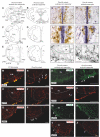
Comparison of the c-fos expression between acidosis-responder and acidosis-non-responder Phox2b mutant preparations under etonogestrel. Drawings of representative sections from the medulla oblongata at the caudal level (with inferior olives; A, B), intermediate level (between inferior olives and the facial nucleus; C, D) and rostral level (with the facial nucleus; E, F) illustrating the c-FOS distribution in non-acidosis responder (A, C, E) and acidosis-responder (B, D, F) Phox2b mutants under etonogestrel. Photomicrographs of c-FOS/5-HT immunoreactivities in the caudal ROb of acidosis-non responder (G, I) and acidosis-responder (H, J) Phox2b mutants. Photomicrographs in (I, J) correspond to enlargements of the outlined area in (G, H); note the greater number of dual-labeled c-FOS/5-HT neurons (*) in acidosis-responder than in non-acidosis responder Phox2b mutants. Photomicrographs of c-FOS immunoreactivity in the ventral medullary surface, ventromedial to the facial nucleus in the pf-RTN (K, L) showing that the number of c-FOS positive neurons (solid black arrows) was greater in acidosis-responder than in non-acidosis-responder Phox2b mutants. Photomicrographs of immunofluorescence detection for PHOX2B in wildtype littermates (M, O, Q) and Phox2b mutants (N, P, R) in the ventral medullary surface, just below the caudal edge of the facial nucleus, in the cRTN (M, N), ventromedial to the facial nucleus, in the pf-RTN (O, P) and ventrolateral below the facial nucleus, in the pFRG (Q, R). In wildtype littermates, PHOX2B neurons are distributed in all three delimitations with the highest number of neurons in the cRTN as already described (5). In Phox2b mutants, the number of neurons was drastically reduced but some residual cells were still present as previously described (hollow white arrows) (6). Dual c-FOS (S, T, W, X) and PHOX2B (U, V, Y, Z) detections by immunofluorescence under the ventromedial part of the RTN (pf-RTN) in acidosis-non-responder (S, U) and acidosis-responder (T, V) Phox2b mutants and under the pFRG in acidosis-non-responder (W, Y) and acidosis-responder (X, Z) Phox2b mutants; note the presence of c-FOS positive (solid white arrows) but PHOX2B negative cells in acidosis-responder Phox2b mutants. Scale bar = 200 μm (A–F), 100 µm (M–R), 50 μm (G, H), and 20 µm (I, J, K, L, S, T, U, V). 7N, facial nucleus; 10N, dorsal motor nucleus of the vagus; 12N, hypoglossal nucleus; Amb, ambiguus nucleus; AP, area postrema; c/mNTS, commissural and median parts of the nucleus of the tractus solitarius; IO, inferior olives; VLM, ventrolateral medullary reticular nucleus; pFRG, parafacial respiratory group; pyr, pyramidal tract; ROb, raphe obscurus nucleus (caudal part—cROb, from pyramidal decussation to rostral edge of the inferior olive and rostral part—rROb, from rostral edge of the inferior olives to rostral edge of the facial nucleus); RPa, raphe pallidus nucleus (caudal part—cRPa, from pyramidal decussation to rostral edge of the inferior olive and rostral part—rRPa, from rostral edge of the inferior olives to rostral edge of the facial nucleus); RTN, retrotrapezoid nucleus (parafacial RTN—pf-RTN, in ventromedial position under the facial nucleus and caudal RTN—cRTN, immediately to the caudal edge of the facial nucleus); sp5, spinal trigeminal tract; Sp5, spinal trigeminal interpolaris nucleus; vlNTS, ventrolateral part of the nucleus of the tractus solitaries; VLM, ventrolateral medullary reticular nucleus; WT, wildtype. Note that the blue staining along the medial axis in (G–J) photomicrographs corresponds to background observed when c-FOS labeling was revealed with Ni-concentrated DAB solution so that the contrast between c-FOS-positive nuclei and 5-HT-positive soma could be clearly distinguished.
Figure 5
Figure 5
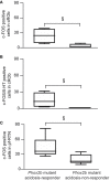
Graphical representation of the increase in number of c-FOS or c-FOS/5-HT positive cells in ROb and pf-RTN in Phox2b mutant acidosis-responder compared to acidosis non-responder under metabolic acidosis with etonogestrel. Box and whisker (median [Q1; Q3] and minimum and maximum values) showing the c-FOS (A, C) or c-FOS/5-HT (B) positive cells in the cROB (A, B) and pf-RTN (C). (§p < 0.05; Mann & Whitney test). cROb (caudal part of the raphe obscurus nucleus; from pyramidal decussation to rostral edge of the inferior olive); pf-RTN (parafacial retrotrapezoid nucleus; in ventromedial position under the facial nucleus).
Figure 6
Figure 6
Effect of an increase in 5-HT signaling by fluoxetine on the respiratory response to metabolic acidosis under exposure to etonogestrel in OF1 mice. (A), Scatter plots with a superimposed box and whisker plot (median [Q1; Q3]) showing percentage of change of fR under 6.25 (n=10), 12.5 (n=11), 25 (n=10), 50 (n=4) and 100 µM (n=4) of fluoxetine. **p < 0.01 indicates a significant change in fR relative to pre-fluoxetine values (paired t-test or Wilcoxon matched pairs signed rank test depending on the normality of data distribution). (B–G), Individual phrenic activity traces (fourth cervical ventral nerve root, C4) and integrated C4 activity from medullary-spinal cord from OF1 mice in normal-pH (B–D; pre-metabolic acidosis) and metabolic acidosis (E–G) conditions, under etonogestrel (B, E), fluoxetine (C, F), or etonogestrel/fluoxetine (D, G) exposure. (H), Median with interquartile range [Q1; Q3] illustrating the respiratory-like rhythm (respiratory frequency, fR) during the metabolic acidosis challenge per 5-min window under etonogestrel (red circles, n=18), fluoxetine (orange circles, n=11), and etonogestrel/fluoxetine (purple circles, n=29) exposure. Under etonogestrel/fluoxetine, preparations displayed a powerful potentiation of the respiratory response to metabolic acidosis compared to etonogestrel or fluoxetine alone. *Indicates a significant difference in fR relative to pre-metabolic acidosis values (**p < 0.01, ***p < 0.001, ****p < 0.0001; one-way analysis of variance or Friedman test followed by Benjamini, Krieger and Yekutieli’s multiple comparison test). §Indicates a significant difference between etonogestrel/fluoxetine preparations and etonogestrel or fluoxetine preparations alone (§§ p < 0.05; §§p < 0.01 two-way analysis of variance test followed by Benjamini, Krieger and Yekutieli’s multiple comparison test). ∫C4: integrated activity of C4 ventral nerve root; C4: electrical activity of the C4 ventral nerve root.
Figure 7
Figure 7
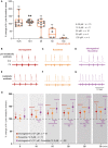
Effect of an increase in 5-HT signaling by fluoxetine in Phox2b mutant mice. Scatter plots with a superimposed box and whisker plot (median [Q1; Q3]) showing percentage of change of fR under fluoxetine in Phox2b mutants (A) and wildtype littermates (B). In Phox2b mutants, 3.125 (n=14), 6.25 (n=12) and 12.5 µM (n=10) of fluoxetine. In wildtype littermates, 3.125 (n=15), 6.25 (n=16) and 12.5 µM (n=15) of fluoxetine. * p < 0.05 indicates a significant change in fR relative to pre-fluoxetine values (paired t-test or Wilcoxon matched pairs signed rank test depending on the normality of data distribution). (C–E), Individual phrenic activity traces (fourth cervical ventral nerve root, C4) and integrated C4 activity from medullary-spinal cord from Phox2b mutant mice in metabolic acidosis under drug-free (C, n=23), etonogestrel (D, n=39) and etonogestrel/fluoxetine (E, n=14) exposure. (F), Scatter plot with surperimposed median [Q1; Q3] showing the respiratory-like rhythm (respiratory frequency, fR) during the last five minutes of metabolic acidosis in percentage of pre-metabolic acidosis. In the presence of fluoxetine, the restoration of the respiratory response to metabolic acidosis induced by etonogestrel in Phox2b mutant mice was abolished. *Indicates a significant change in fR relative to pre-metabolic acidosis values (*p < 0.05; paired t-test). §Indicates a significant difference between the 3 conditions (§p < 0.05; Kruskal-Wallis test followed by Benjamini, Krieger and Yekutieli’s multiple comparison test). ∫C4, integrated activity of C4 ventral nerve root; C4, electrical activity of the C4 ventral nerve root. Yellow squares and pink circles represent respectively male and female Phox2b mutant mice acidosis-responders (+10% above pre-metabolic values) and green squares and grey circles represent respectively male and female Phox2b mutant mice acidosis-non-responders under, drug-free, etonogestrel and etonogestrel/fluoxetine, respectively.
Figure 8
Figure 8
Ultra-high-performance liquid chromatography analysis of 5-HT contents and its related compounds, 5-HTP and 5-HIAA, in the medulla oblongata of Phox2b mutants, wildtype littermates and OF1 mice. (A), Example of a chromatogram of medulla oblongata obtained from Phox2b mutants (blue; n=8), wildtype littermates (pink; n=8) and OF1 mice (purple; n=10) with indicated peaks of serotonin (5-HT), its precursor 5-hydroxytryptophan (5-HTP) and its metabolite 5-hydroxyindole acetic acid (5-HIAA). (B), Scatter plots showing 5-HT quantity (g of 5-HT per medulla) with superimposed mean ± standard error of the mean. (C, D), Scatter plots showing, respectively, 5-HT/5-HTP and 5-HIAA/5-HT ratio with median [Q1; Q3] superimposed in OF1 (purple filled circles), wildtype littermates (pink filled circles) and Phox2b mutants (blue filled circles). * Indicates a significant difference between OF1, Phox2b mutants and wildtype littermates (**p < 0.01 and ***p < 0.001; one-way analysis of variance or Kruskal-Wallis test followed by Benjamini, Krieger and Yekutieli’s multiple comparison test) ns, non significant.

Similar articles

Cited by

References

    1. Trang H, Samuels M, Ceccherini I, Frerick M, Garcia-Teresa MA, Peters J, et al. . Guidelines for diagnosis and management of congenital central hypoventilation syndrome. Orphanet J Rare Dis (2020) 15:252. doi: 10.1186/s13023-020-01460-2 - DOI - PMC - PubMed
    1. Amiel J, Laudier B, Ttie-Bitach T, Trang H, De PL, Gener B, et al. . Polyalanine expansion and frameshift mutations of the paired-like homeobox gene PHOX2B in congenital central hypoventilation syndrome. Nat Genet (2003) 33:459–61. doi: 10.1038/ng1130 - DOI - PubMed
    1. Amiel J, Dubreuil V, Ramanantsoa N, Fortin G, Gallego J, Brunet JF, et al. . PHOX2B in respiratory control: lessons from congenital central hypoventilation syndrome and its mouse models. Respir Physiol Neurobiol (2009) 168:125–32. doi: 10.1016/j.resp.2009.03.005 - DOI - PubMed
    1. Weese-Mayer DE, Rand CM, Zhou A, Carroll MS, Hunt CE. Congenital central hypoventilation syndrome: a bedside-to-bench success story for advancing early diagnosis and treatment and improved survival and quality of life. Pediatr Res (2017) 81:192–201. doi: 10.1038/pr.2016.196 - DOI - PubMed
    1. Dubreuil V, Ramanantsoa N, Trochet D, Vaubourg V, Amiel J, Gallego J, et al. . A human mutation in Phox2b causes lack of CO2 chemosensitivity, fatal central apnea, and specific loss of parafacial neurons. Proc Natl Acad Sci U.S.A. (2008) 105:1067–72. doi: 10.1073/pnas.0709115105 - DOI - PMC - PubMed

Publication types

Supplementary concepts

LinkOut - more resources