

Hickam's Dictum Incarnate: A Case of Simultaneous Left-Sided Urolithiasis and Ruptured Iliac Artery Aneurysm
- PMID: 36896276
- PMCID: PMC9979919
- DOI: 10.24908/pocus.v7i1.15020
Hickam's Dictum Incarnate: A Case of Simultaneous Left-Sided Urolithiasis and Ruptured Iliac Artery Aneurysm
Abstract
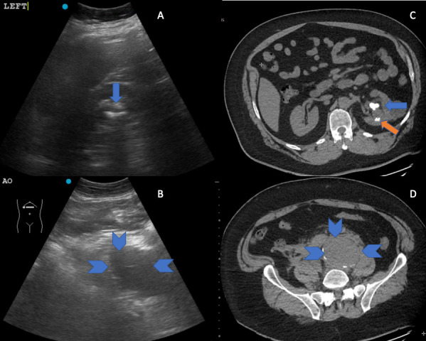
A 51-year-old man with a history of nephrolithiasis presented to the Emergency Department after a sudden onset of left-sided groin pain and syncope. At presentation, he described his pain as similar to prior renal colic episodes. At his initial assessment, point of care ultrasound (POCUS) was used, which revealed findings consistent with obstructive renal stones, as well as a substantially enlarged left iliac artery. Computed tomography (CT) imaging confirmed the comorbid diagnoses of left-sided urolithiasis and a ruptured isolated left iliac artery aneurysm. POCUS facilitated expedited definitive imaging and operative management. This case highlights the importance of performing related POCUS studies in reducing anchoring and premature closure bias.
Keywords: abdominal aortic aneurysm; iliac artery aneurysm; nephrolithiasis; point of care ultrasound; ultrasound; urolithiasis.
Copyright (c) 2022 Melissa Bouwsema, Colin Bell.
Conflict of interest statement
The authors have no conflicts of interest to declare.
Figures
References
-
- Physicians American College of Emergency. Emergency Ultrasound Imaging Criteria Compendium. Annals of Emergency Medicine. 2006;48(4):487–510. - PubMed
-
- Lewiss R E, Strony Robert J, Jones Robert A. Practical Guide to Critical Ultrasound. American College of Emergency Physicians; Dallas, Texas: 2018. p. 221.
-
- Tayal V S, Graf C D, Gibbs M A. Prospective study of accuracy and outcome of emergency ultrasound for abdominal aortic aneurysm over two years. Academic Emergency Medicine. 2003;10(8):867–871. - PubMed
-
- Dix F P, Titi M, H Al-Khaffaf. The isolated internal iliac artery aneurysm-a review. European Journal of Vascular and Endovascular Surgery. 2005;30(2):119–129. - PubMed
-
- Bhatt S, Dogra V S. Catastrophes of abdominal aorta: sonographic evaluation. Ultrasound Clinics. 2008;3(1):83–91.
Publication types
LinkOut - more resources
Full Text Sources