

Mesh erosion into the colon following repair of parastomal hernia: A case report
- PMID: 36896303
- PMCID: PMC9988641
- DOI: 10.4240/wjgs.v15.i2.294
Mesh erosion into the colon following repair of parastomal hernia: A case report
Abstract
Background: In recent years, mesh has become a standard repair method for parastomal hernia surgery due to its low recurrence rate and low postoperative pain. However, using mesh to repair parastomal hernias also carries potential dangers. One of these dangers is mesh erosion, a rare but serious complication following hernia surgery, particularly parastomal hernia surgery, and has attracted the attention of surgeons in recent years.
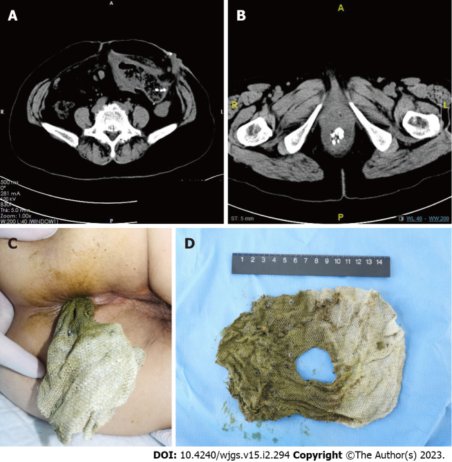
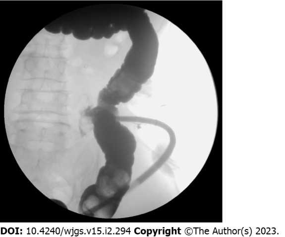
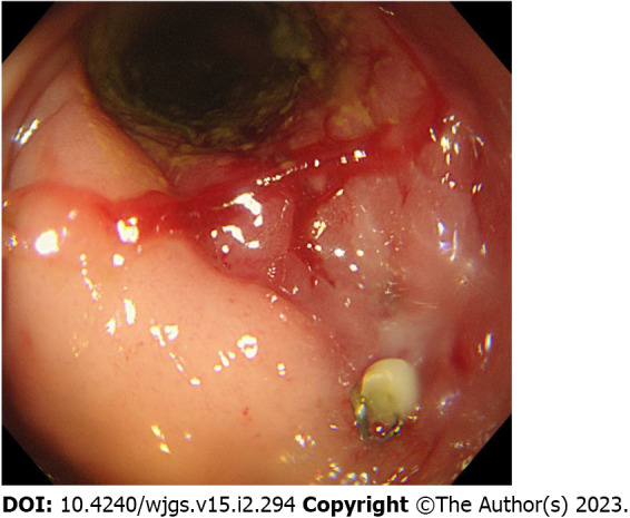
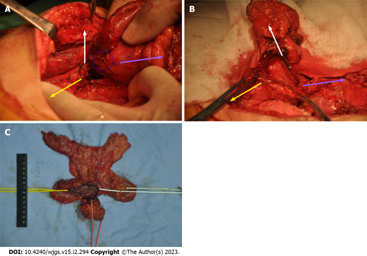
Case summary: Herein, we report the case of a 67-year-old woman with mesh erosion after parastomal hernia surgery. The patient, who underwent parastomal hernia repair surgery 3 years prior, presented to the surgery clinic with a complaint of chronic abdominal pain upon resuming defecation through the anus. Three months later, a portion of the mesh was excreted from the patient's anus and was removed by a doctor. Imaging revealed that the patient's colon had formed a t-branch tube structure, which was formed by the mesh erosion. The surgery reconstructed the structure of the colon and eliminated potential bowel perforation.
Conclusion: Surgeons should consider mesh erosion since it has an insidious development and is difficult to diagnose at the early stage.
Keywords: Case report; Intestinal fistula; Intestinal internal fistula; Mesh erosion; Mesh migration; Parastomal hernia.
©The Author(s) 2023. Published by Baishideng Publishing Group Inc. All rights reserved.
Conflict of interest statement
Conflict-of-interest statement: All the authors report no relevant conflicts of interest for this article.
Figures
Similar articles
-
Safety and outcome of use of nonabsorbable mesh for repair of fascial defects in the presence of open bowel.Dis Colon Rectum. 2003 Aug;46(8):1118-23. doi: 10.1007/s10350-004-7290-x. Dis Colon Rectum. 2003. PMID: 12907910
-
Parastomal hernias -- clinical study of therapeutic strategies.Chirurgia (Bucur). 2014 Mar-Apr;109(2):179-84. Chirurgia (Bucur). 2014. PMID: 24742407 Clinical Trial.
-
Single center experience with the modified retromuscular Sugarbaker technique for parastomal hernia repair.Hernia. 2017 Dec;21(6):941-949. doi: 10.1007/s10029-017-1644-5. Epub 2017 Aug 24. Hernia. 2017. PMID: 28840354
-
Chronic abdominal pain after ventral hernia due to mesh migration and erosion into the sigmoid colon from a distant site: a case report and review of literature.Hernia. 2015 Oct;19(5):849-52. doi: 10.1007/s10029-013-1182-8. Epub 2013 Nov 20. Hernia. 2015. PMID: 24253380 Review.
-
Parastomal hernia: complications of extra-peritoneal onlay mesh placement.Hernia. 2009 Oct;13(5):487-90. doi: 10.1007/s10029-009-0494-1. Epub 2009 Mar 26. Hernia. 2009. PMID: 19322626 Review.
Cited by
-
Use of prophylactic mesh to prevent parastomal hernia formation: a systematic review, meta-analysis and network meta-analysis.Hernia. 2024 Nov 18;29(1):22. doi: 10.1007/s10029-024-03219-1. Hernia. 2024. PMID: 39556272
-
Robotic Retromuscular (Recurrent) Parastomal Hernia Repair (r-Pauli-Repair) With Synthetically Reinforced Biological Mesh; Technique, Early Experience, and Short-Term Follow-Up.J Abdom Wall Surg. 2023 Dec 13;2:12059. doi: 10.3389/jaws.2023.12059. eCollection 2023. J Abdom Wall Surg. 2023. PMID: 38312416 Free PMC article.
-
Are Surgeons Going to Be Left Holding the Bag? Incisional Hernia Repair and Intra-Peritoneal Non-Absorbable Mesh Implant Complications.J Clin Med. 2024 Feb 9;13(4):1005. doi: 10.3390/jcm13041005. J Clin Med. 2024. PMID: 38398318 Free PMC article. Review.
References
-
- Antoniou SA, Agresta F, Garcia Alamino JM, Berger D, Berrevoet F, Brandsma HT, Bury K, Conze J, Cuccurullo D, Dietz UA, Fortelny RH, Frei-Lanter C, Hansson B, Helgstrand F, Hotouras A, Jänes A, Kroese LF, Lambrecht JR, Kyle-Leinhase I, López-Cano M, Maggiori L, Mandalà V, Miserez M, Montgomery A, Morales-Conde S, Prudhomme M, Rautio T, Smart N, Śmietański M, Szczepkowski M, Stabilini C, Muysoms FE. European Hernia Society guidelines on prevention and treatment of parastomal hernias. Hernia . 2018;22:183–198. - PubMed
-
- Hansson BM, Slater NJ, van der Velden AS, Groenewoud HM, Buyne OR, de Hingh IH, Bleichrodt RP. Surgical techniques for parastomal hernia repair: a systematic review of the literature. Ann Surg . 2012;255:685–695. - PubMed
-
- Cunningham HB, Kukreja S, Huerta S. Mesh migration into an inguinal hernia sac following a laparoscopic umbilical hernia repair. Hernia . 2018;22:715–720. - PubMed
-
- Jeans S, Williams GL, Stephenson BM. Migration after open mesh plug inguinal hernioplasty: a review of the literature. Am Surg . 2007;73:207–209. - PubMed
-
- Hamouda A, Kennedy J, Grant N, Nigam A, Karanjia N. Mesh erosion into the urinary bladder following laparoscopic inguinal hernia repair; is this the tip of the iceberg? Hernia . 2010;14:317–319. - PubMed
Publication types
LinkOut - more resources
Full Text Sources
