. 2023 Mar 25;55(3):508-517.
doi: 10.3724/abbs.2023031.

Fun30 nucleosome remodeller regulates white-to-opaque switching in Candida albicans

Affiliations

Fun30 nucleosome remodeller regulates white-to-opaque switching in Candida albicans

Ning Gao et al. Acta Biochim Biophys Sin (Shanghai). .

Abstract

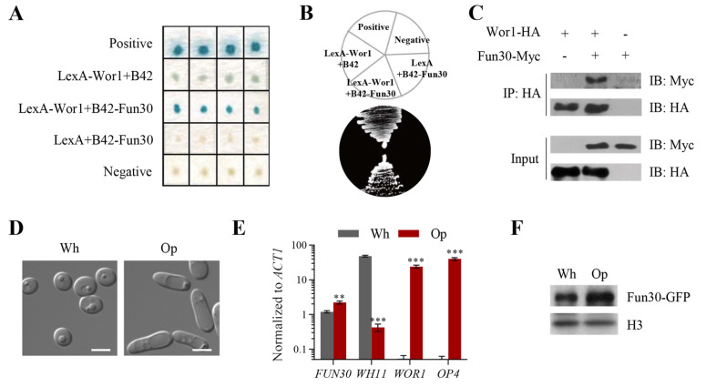
Candida albicans ( C. albicans) is an opportunistic pathogen in humans and possesses a white-opaque heritable switching system. Wor1 is a master regulator of white-opaque switching and is essential for opaque cell formation in C. albicans. However, the regulatory network of Wor1 in white-opaque switching is still vague. In this study, we obtain a series of Wor1-interacting proteins using LexA-Wor1 as bait. Among these proteins, function unknown now 30 (Fun30) interacts with Wor1 in vitro and in vivo. Fun30 expression is upregulated in opaque cells at the transcriptional and protein levels. Loss of FUN30 attenuates white-to-opaque switching, while ectopic expression of FUN30 significantly increases white-to-opaque switching in an ATPase activity-dependent manner. Furthermore, FUN30 upregulation is dependent on CO 2; loss of FLO8, a key CO 2-sensing transcriptional regulator, abolishes FUN30 upregulation. Interestingly, deletion of FUN30 affects the WOR1 expression regulation feedback loop. Thus, our results indicate that the chromatin remodeller Fun30 interacts with Wor1 and is required for WOR1 expression and opaque cell formation.

Keywords: Fun30; Wor1; chromatin remodeller; white-opaque switching.


Conflict of interest statement

The authors declare that they have no conflict of interest.

Figures

None
Figure 1
Fun30 is a novel Wor1-interacting protein and is upregulated in opaque cells (A,B) Yeast two-hybrid screen. EGY48 (p8op-lacZ) was cotransformed with plasmids as described in the Materials and Methods section. The protein-protein interactions were analyzed by (A) a β-galactosidase activity assay using X-gal as the substrate and (B) a growth ability assay on SC Gal His – Trp – Leu – plates. Plasmid pSH17-4 containing a LexA-AD fusion was used as the positive control, and pRFHM1 containing a LexA-human lamin C fusion was used as the negative control. (C) Co-IP assay. Plasmid pPR673-FUN30 containing Fun30-Myc was introduced into C. albicans strain CYM2 carrying Wor1-HA from the WOR1 promoter. Protein lysates from opaque cells were immunoprecipitated with anti-Myc antibody and immunoblotted with HA or a Myc probe. The cell lysates were also analyzed by immunoblotting with peroxidase-conjugated anti-HA or anti-Myc as the input control. (D) Representative cell morphology from the white (Wh) or opaque (Op) colonies of the WT strain (JYC5+pACT1) on a YPD plate incubated at 22°C for 5 days. Scale bar: 5 μm. (E) Expressions of FUN30, WH11, WOR1, and OP4 in WT white and opaque cells from (D) calculated by quantitative real-time PCR (qRT-PCR) analysis. The signal obtained from ACT1 mRNA was used as a reference control for normalization. Data are representative of at least three independent experiments and shown as the mean±SD, each with similar results. * P<0.05, ** P<0.01, *** P<0.001, unpaired Student’s t test. (F) Western blot analysis was used to detect GFP-tagged Fun30 in white and opaque cells. White or opaque cells containing endogenously expressed Fun30-GFP (JYC5+pFUN30-GFP) were cultured to log phase in YPD at 22°C; cell lysates were immunoblotted with anti-GFP antibody, and histone H3 was used as a loading control. Data represent at least three independent experiments, each with similar results.
None
Figure 2
Fun30 is a Swr1-like protein belonging to the Snf2 subfamily and localizes to the nucleus (A) A schematic diagram of Fun30 and other Snf2 subfamily members in C. albicans (Ca) and S. cerevisiae (Sc), including Fun30, Swr1, Ino80 and Snf2. HSA domains (yellow) and ATPase domains (blue). The bromo domain is specific to Snf2. (B) Subcellular localization of Fun30 in C. albicans . Wild-type (a/a) and (a/α) cells expressing the Fun30-GFP reporter gene. GFP was tagged at the C-terminus of Fun30. DAPI staining (blue) shows the nucleus. DIC, differential interference contrast. Scale bar: 5 μm.
None
Figure 3
Fun30 promotes opaque cell formation in an ATPase activity-dependent manner (A) White-to-opaque switching assays. The white cells from WT (JYC5+pACT1) and/or fun30 mutant strains were plated onto YPD or YPG plates and incubated in 20% CO 2 at 22°C. Two thousand cells from each sample were collected, and cell morphology was examined every 2 days; the percentage of opaque cells was then calculated to determine the white-to-opaque switching frequency. Data are representative of at least three independent experiments and shown as the mean± SD. * P<0.05, unpaired Student’s t test. (B) A schematic diagram mapping the region of Fun30 required for opaque formation regulation. The ATPase-N and ATPase-C domains comprise the ATPase domain, which is the catalytic core of ATP hydrolysis and the site of DNA binding. K593R is a point mutation in the ATPase domain that replaces lysine at position 593 (AAA) by arginine (AGA), and this mutation results in the dramatic loss of ATPase activity in C. albicans, as shown by sequence alignment. (C) Expression of FUN30 in WT+Vec, WT+ FUN30, and WT+ FUN30 K593R strains. The white cells from these strains were cultured to log phase in YPD at 22°C and then collected for qRT-PCR analysis. The signal obtained from ACT1 mRNA was used as a reference control for normalization. Data are representative of at least three independent experiments and shown as the mean±SD. * P<0.05, ** P<0.01, unpaired Student’s t test. (D) Overexpression of FUN30 enhanced white-to-opaque switching. White cells of the WT strain JYC5 carrying pBA1, pBA1-FUN30, and pBA1-FUN30-K593R were cultured in YPD, plated onto SCD plates containing phloxine B and incubated at 22°C for 7 days. The colonies were photographed, and the percentage of opaque cells was calculated to determine the white-to-opaque switching frequency (E). Approximately 1000 colonies from each sample were counted per experiment, and cell morphology was examined. Data are representative of at least three independent experiments and shown as the mean±SD. * P<0.05, ** P<0.01, unpaired Student’s t test. ns, no significance.
None
Figure 4
Wor1 controls the recruitment of Fun30 to the promoter of WOR1 and promotes WOR1 expression (A) ChIP-qPCR analysis of Fun30 at the Wor1 binding sites on the WOR1 promoter. The enrichment of Fun30 was detected at five Wor1 binding sites in the WOR1 promoter: –7900, –6100, –5100, –4100, and –2500 [14]; the –350 site was a nonspecific site acting as a negative control [14]. The cells used for the assay were as follows: WT+V (JYC5+pPR673) opaque cells, WT+Fun30-Myc (JYC5+pPR673-FUN30) opaque cells, WT+Fun30-Myc white cells and wor 1 ( URA3 -)+Fun30-Myc white cells. The signal obtained from WT+Fun30-Myc opaque cells at the –350 site was used for normalization. (B) Expression of WOR1 in WT+V and fun30 mutant strains with 20% CO 2 induction. The white cells were spread onto a YPD plate and incubated at 22°C for 2 days in 20% CO 2. The cells were collected for qRT-PCR analysis. The signal obtained from ACT1 mRNA was used as a reference control for normalization. Data are representative of at least three independent experiments and shown as the mean±SD. * P<0.05, ** P<0.01, *** P<0.001, unpaired Student’s t test.
None
Figure 5
FUN30 is upregulated in response to CO 2, dependent on the CO 2-sensing transcription factor Flo8 (A) FUN30 transcription in WT+V (JYC5+pBA1) is elevated in response to CO 2 treatment, as shown by qRT-PCR. (B) Expression of FUN30 in the WT+V and flo8+V strains. Briefly, log-phase white cells were spread onto YPD plates and incubated at 22°C for 2 days in different concentrations of CO 2. The cells were collected for qRT-PCR analysis, as shown in (A) & (B). The signal obtained from ACT1 mRNA was used as a reference control for normalization. Data are representative of at least three independent experiments and shown as the mean±SD. * P<0.05, ** P<0.01, *** P<0.001, unpaired Student’s t test.
None
Figure 6
Model of Fun30-regulated Wor1 interaction and transcriptional regulation during white-to-opaque switching in C. albicans The Fun30 nucleosome remodeller is upregulated upon high CO 2 stimulation in a Flo8-dependent manner. The elevated Fun30 then binds with Wor1 and is recruited to the promoter of WOR1, thereby participating in the positive feedback loop of WOR1 regulation. Thus, Fun30 is an upstream epigenetic factor regulating white-to-opaque switching in C. albicans.

Similar articles

References

    1. Kadosh D. Regulatory mechanisms controlling morphology and pathogenesis in Candida albicans . Curr Opin Microbiol. . 2019;52:27–34. doi: 10.1016/j.mib.2019.04.005. - DOI - PMC - PubMed
    1. Noble SM, Gianetti BA, Witchley JN. Candida albicans cell-type switching and functional plasticity in the mammalian host . Nat Rev Microbiol. . 2017;15:96–108. doi: 10.1038/nrmicro.2016.157. - DOI - PMC - PubMed
    1. Slutsky B, Buffo J, Soll DR. High-frequency switching of colony morphology in Candida albicans . Science. . 1985;230:666–669. doi: 10.1126/science.3901258. - DOI - PubMed
    1. Rikkerink EH, Magee BB, Magee PT. Opaque-white phenotype transition: a programmed morphological transition in Candida albicans . J Bacteriol. . 1988;170:895–899. doi: 10.1128/jb.170.2.895-899.1988. - DOI - PMC - PubMed
    1. Slutsky B, Staebell M, Anderson J, Risen L, Pfaller M, Soll DR. White-opaque transition: a second high-frequency switching system in Candida albicans . J Bacteriol. . 1987;169:189–197. doi: 10.1128/jb.169.1.189-197.1987. - DOI - PMC - PubMed