. 2023 Sep;19(9):2485-2503.
doi: 10.1080/15548627.2023.2186099. Epub 2023 Mar 10.

Phosphorylation of KRT8 (keratin 8) by excessive mechanical load-activated PKN (protein kinase N) impairs autophagosome initiation and contributes to disc degeneration

Affiliations

Phosphorylation of KRT8 (keratin 8) by excessive mechanical load-activated PKN (protein kinase N) impairs autophagosome initiation and contributes to disc degeneration

Di Wang et al. Autophagy. 2023 Sep.

Abstract

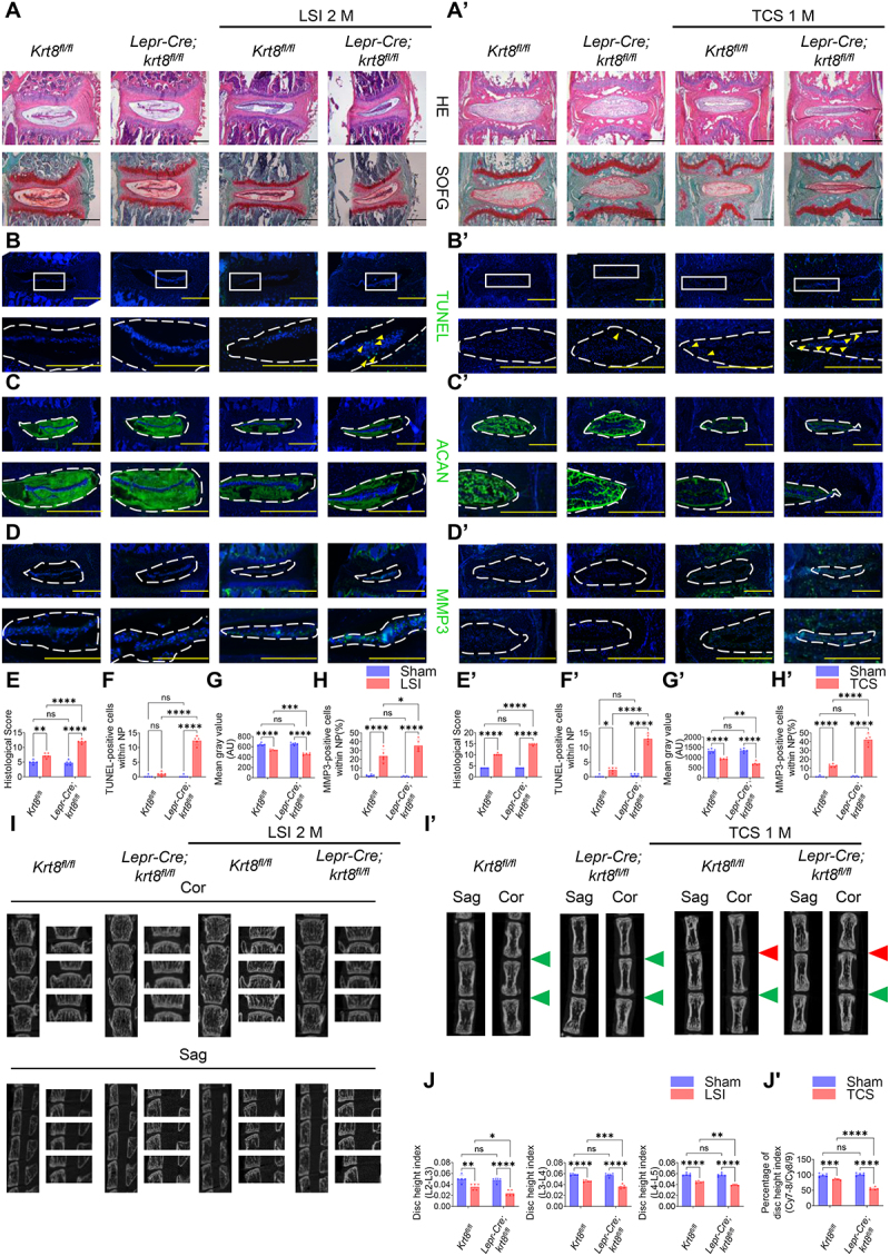
Excessive mechanical load (overloading) is a well-documented pathogenetic factor for many mechano stress-induced pathologies, i.e. intervertebral disc degeneration (IDD). Under overloading, the balance between anabolism and catabolism within nucleus pulposus (NP) cells are badly thrown off, and NP cells undergo apoptosis. However, little is known about how the overloading is transduced to the NP cells and contributes to disc degeneration. The current study shows that conditional knockout of Krt8 (keratin 8) within NP aggravates load-induced IDD in vivo, and overexpression of Krt8 endows NP cells greater resistance to overloading-induced apoptosis and degeneration in vitro. Discovery-driven experiments shows that phosphorylation of KRT8 on Ser43 by overloading activated RHOA-PKN (protein kinase N) impedes trafficking of Golgi resident small GTPase RAB33B, suppresses the autophagosome initiation and contributes to IDD. Overexpression of Krt8 and knockdown of Pkn1 and Pkn2, at an early stage of IDD, ameliorates disc degeneration; yet only knockdown of Pkn1 and Pkn2, when treated at late stage of IDD, shows a therapeutic effect. This study validates a protective role of Krt8 during overloading-induced IDD and demonstrates that targeting overloading activation of PKNs could be a novel and effective approach to mechano stress-induced pathologies with a wider window of therapeutic opportunity.Abbreviations: AAV: adeno-associated virus; AF: anulus fibrosus; ANOVA: analysis of variance; ATG: autophagy related; BSA: bovine serum albumin; cDNA: complementary deoxyribonucleic acid; CEP: cartilaginous endplates; CHX: cycloheximide; cKO: conditional knockout; Cor: coronal plane; CT: computed tomography; Cy: coccygeal vertebra; D: aspartic acid; DEG: differentially expressed gene; DHI: disc height index; DIBA: dot immunobinding assay; dUTP: 2'-deoxyuridine 5'-triphosphate; ECM: extracellular matrix; EDTA: ethylene diamine tetraacetic acid; ER: endoplasmic reticulum; FBS: fetal bovine serum; GAPDH: glyceraldehyde-3-phosphate dehydrogenase; GPS: group-based prediction system; GSEA: gene set enrichment analysis; GTP: guanosine triphosphate; HE: hematoxylin-eosin; HRP: horseradish peroxidase; IDD: intervertebral disc degeneration; IF: immunofluorescence staining; IL1: interleukin 1; IVD: intervertebral disc; KEGG: Kyoto encyclopedia of genes and genomes; KRT8: keratin 8; KD: knockdown; KO: knockout; L: lumbar vertebra; LBP: low back pain; LC/MS: liquid chromatograph mass spectrometer; LSI: mouse lumbar instability model; MAP1LC3/LC3: microtubule associated protein 1 light chain 3; MMP3: matrix metallopeptidase 3; MRI: nuclear magnetic resonance imaging; NC: negative control; NP: nucleus pulposus; PBS: phosphate-buffered saline; PE: p-phycoerythrin; PFA: paraformaldehyde; PI: propidium iodide; PKN: protein kinase N; OE: overexpression; PTM: post translational modification; PVDF: polyvinylidene fluoride; qPCR: quantitative reverse-transcriptase polymerase chain reaction; RHOA: ras homolog family member A; RIPA: radio immunoprecipitation assay; RNA: ribonucleic acid; ROS: reactive oxygen species; RT: room temperature; TCM: rat tail compression-induced IDD model; TCS: mouse tail suturing compressive model; S: serine; Sag: sagittal plane; SD rats: Sprague-Dawley rats; shRNA: short hairpin RNA; siRNA: small interfering RNA; SOFG: safranin O-fast green; SQSTM1: sequestosome 1; TUNEL: terminal deoxynucleotidyl transferase dUTP nick end labeling; VG/ml: viral genomes per milliliter; WCL: whole cell lysate.

Keywords: Autophagosome initiation; Protein kinase N; intervertebral disc degeneration; keratin 8; phosphorylation.


Conflict of interest statement

No potential conflict of interest was reported by the authors.

Figures

Figure 1.
Figure 1.
Conditional knockout of Krt8 within NP aggravated load-induced disc degeneration in vivo. Krt8fl/fl and Lepr-Cre;krt8fl/fl mice were subjected to LSI or TCS. (A) HE and SOFG staining of L4-L5 disc sections. (A’) HE and SOFG staining of Cy7-Cy8 disc sections. (B) TUNEL assay of L4-L5 disc sections. (B‘) TUNEL assay of Cy7-Cy8 disc sections. (C) Immunofluorescence staining of ACAN of L4-L5 disc sections. (C’) Immunofluorescence staining of ACAN of Cy7-Cy8 disc sections. (D) Immunofluorescence staining of MMP3 of L4-L5 disc sections. (D’) Immunofluorescence staining of MMP3 of Cy7-Cy8 disc sections. (E) Histological score of Fig1A. (E’) Histological score of Fig1A’. (F) Quantification of TUNEL-positive cells of Fig1B. (F’) Quantification of TUNEL-positive cells of Fig1B’. (G) Fluorescence intensity of ACAN of Fig1c. (G’) Fluorescence intensity of ACAN of Fig1c’. (H) Quantification of MMP3-positive cells of Fig1D. (H’) Quantification of MMP3-positive cells of Fig1D’. (I) Micro CT analysis of L2-L5 vertebras. (Cor: coronal plane; Sag: sagittal plane) (I’) Micro CT analysis of Cy7-Cy8 vertebras. (Cor: coronal plane; sag: Sagittal plane) (J) Quantification of DHI of L2-L3, L3-L4, L4-L5 discs. (J’) Quantification of percentage of DHI (Cy7–8÷cy8–9). n = 5; *: p<0.05; **: p<0.01, ***: p<0.005, ****: p<0.0001 ns: not statistically significant; Scale bar: 50 μm, the yellow triangle indicates TUNEL-positive cells. For all the statistical analyses in this figure, significance was determined by Two-way ANOVA followed by Tukey’s multiple comparisons test, and results were shown as mean ± S.D.
Figure 2.
Figure 2.
Loss of Krt8 predisposed NP cells to, and overexpression of Krt8 endowed NP cells with greater resistance to, excessive mechanical load-induced apoptosis and degeneration in vitro. Primary rat NP cells were subjected to in vitro compression model, siRNA-mediated knockdown of Krt8 or adenovirus-mediated overexpression of Krt8 and Krt18 were performed as indicated. (A) Representative images of immunofluorescence staining of KRT8 of NP cells. (B and C) the transcription of Krt8, Acan, Mmp3, Il6 from indicated groups, as determined by qPCR. (D and E) the protein level of KRT8, cleaved CASP3, IL6, MMP3, ACAN from indicated groups, as determined by western blot. (F-H), and (I) Apoptosis of indicated groups evaluated by flowcytometry and its’ quantifications. NC: negative control; n = 3; *: p<0.05; **: p<0.01, ***: p<0.005, ****: p<0.0001 ns: not statistically significant; Scale bar: 20 μm. For all the statistical analyses in this figure, significance was determined by One-way ANOVA followed by Tukey’s multiple comparisons test, and results were shown as mean ± S.D.
Figure 3.
Figure 3.
A damped autophagosome initiation, caused by impeded trafficking of Golgi resident small GTPase RAB33B, was responsible for the detrimental effect of loss of Krt8 on NP cells under excessive mechanical load. Primary rat NP cells were subjected to in vitro compression model, siKrt8 or overexpression of Krt8 and Krt18 were performed as indicated. (A-D) Representative images of immunofluorescence staining of LC3B of NP cells and their quantifications. (E and F) the protein level of LC3B, SQSTM1 from indicated groups. (G). The transcription of Rab33b form indicated groups, as determined by qPCR. (H) the transcription of Rab33b form indicated groups, as determined by mRNA sequencing. (I and J) the protein level of RAB33B from indicated groups, as determined by western blot. (K and L) ATG16L1 bound RAB33B, KRT8 bound RAB33B, Golgi localized RAB33B, ER localized RAB33B and RAB33B from whole cell lysate (WCL) from indicated groups, as determined by western blot. NC: negative control; n = 3; *: p<0.05; **: p<0.01, ***: p<0.005, ****: p<0.0001 ns: not statistically significant; Scale bar: 20 μm. For all the statistical analyses in this figure, significance was determined by One-way ANOVA followed by Tukey’s multiple comparisons test, and results were shown as mean ± S.D.
Figure 4.
Figure 4.
The phosphorylation of KRT8 on Ser43 impeded the trafficking of RAB33B from Golgi to autophagosome through trapping RAB33B with p-Ser43 KRT8, and mechano stress-induced activation of RHOA-PKN was responsible for the phosphorylation of KRT8 on Ser43 under excessive mechanical load. Primary rat NP cells were subjected to in vitro compression for indicated time. (A) the phosphorylation of KRT8, KRT8 bound RAB33B, ATG16L1 bound RBA33B, and KRT8, RAB33B, ATG16L1 protein from WCL of indicated groups were determined by western blot. Primary rat NP cells were subjected to in vitro compression for 48 h, accompanied by overexpression of wild-type Krt8 or mutant Krt8 as indicated. (B) KRT8 bound RAB33B, ATG16L1 bound RBA33B, and KRT8, RAB33B, ATG16L1 protein from WCL of indicated groups were determined by western blot. Primary rat NP cells were subjected to in vitro compression for 48 h, accompanied by siRNA-mediated knockdown of predicted protein kinases. (C) the phosphorylation of KRT8, KRT8 bound RAB33B and ATG16L1 bound RBA33B, of indicated groups were determined by western blot. (D) the quantification of apoptotic cells from indicated groups, determined by flowcytometry. Primary rat NP cells were subjected to in vitro compression for 48 h, accompanied by siRNA-mediated knockdown of Pkn1 and Pkn2 or not. (E) the protein level of cleaved CASP3, IL6, MMP3 and ACAN from indicated groups, as determined by western blot. Primary rat NP cells were subjected to in vitro compression for indicated time period or pressure. (F and G) the activation of PKN1 and PKN2, revealed by the phosphorylation of PKN1 and PKN2, was determine by western blot. (H) GSEA analysis showed an activation of R-RNO-5625740 after 1.0 MPa 24 h compression. Primary rat NP cells were subjected to in vitro compression for indicated period of time, accompanied by treatment of RHOA inhibitor C3 transferase (50 nmol/l) or not. (I) the activation of PKN1 and PKN2, revealed by the phosphorylation of PKN1 and PKN2, and the phosphorylation of KRT8 on Ser43, was determine by western blot. (J) KRT8 bound RAB33B and ATG16L1 bound RAB33B were determined by western blot. n = 3; *: p<0.05; **: p<0.01, ***: p<0.005, ****: p<0.0001 ns: not statistically significant. For all the statistical analyses in this figure, significance was determined by One-way ANOVA followed by Tukey’s multiple comparisons test, and results were shown as mean ± S.D.
Figure 5.
Figure 5.
AAV5-mediated overexpression of Krt8 as well as shRNA-mediated knockdown of Pkn1 and Pkn2, at early stage of TCM, ameliorated rat tail disc degeneration. Eight-week-old male SD rats were subjected to TCM. In this experiment, AAV or shRNA was injected into the NP of the Cy7-Cy8 IVD, immediately after the TCM surgery. (A and B) Summary of the design of the current study. (C) the protein level of KRT8, cleaved CASP3, ACAN, MMP3, and IL6 from indicated groups were determined by western blot. (D and E) HE, SOFG staining and histological score of Cy7-Cy8 disc sections. (F and G) MRI analysis, Pfirrmann grading and its’ quantification of coccygeal discs. (H and I) X-ray analysis, and quantification of percentage of DHI (Cy7–8/Cy8–9) of coccygeal discs. n = 5; *: p<0.05; **: p<0.01, ***: p<0.005, ****: p<0.0001 ns: not statistically significant; Scale bar: 500 μm, the red arrow indicates compression apparats puncture points; the green triangle indicates control discs; the red triangle indicates degenerated discs; and the yellow triangle indicates AAV or shRNA injected discs. For all the statistical analyses in this figure, significance was determined by One-way ANOVA followed by Tukey’s multiple comparisons test, and results were shown as mean ± S.D.
Figure 6.
Figure 6.
The therapeutical effect of AAV-mediated knockdown of Pkn1 and Pkn2 or overexpression of Krt8 and Krt18 at late stage of compression-induced disc degeneration model. 8-week-old male SD rats were subjected to TCM. In this experiment, AAV or shRNA was injected into the NP of the Cy7-Cy8 IVD, 6 weeks after the TCM surgery. (A and B) Summary of the design of the current study. (C) the protein level of KRT8, cleaved CASP3, ACAN, MMP3, and IL6 from indicated groups were determined by western blot. (D and E) HE, SOFG staining and histological score of Cy7-Cy8 disc sections. (F and G) MRI analysis, Pfirrmann grading and its’ quantification of coccygeal discs. (H and I) X-ray analysis, and quantification of percentage of DHI (Cy7–8/Cy8/9) of coccygeal discs. n = 5; *: p<0.05; **: p<0.01, ***: p<0.005, ****: p<0.0001 ns: not statistically significant; Scale bar: 500 μm, the red arrow indicates compression apparats puncture points; the green triangle indicates control discs; the red triangle indicates degenerated discs; and the yellow triangle indicates AAV or shRNA injected discs. For all the statistical analyses in this figure, significance was determined by One-way ANOVA followed by Tukey’s multiple comparisons test, and results were shown as mean ± S.D.
Figure 7.
Figure 7.
Schematic diagram of the current study. Phosphorylation of Ser43 on KRT8 by excessive mechanical load activated RHOA-PKN impedes the trafficking of Golgi resident protein RAB33B, by trapping RABB33B with p-Ser43 KRT8, impairs autophagosome initiation and contributes to disc degeneration.

References

    1. Hoy D, March L, Brooks P, et al. The global burden of low back pain: estimates from the global burden of disease 2010 study. Ann Rheum Dis. 2014;73:968–974. - PubMed
    1. Gianola S, Bargeri S, Del Castillo G, et al. Effectiveness of treatments for acute and subacute mechanical non-specific low back pain: a systematic review with network meta-analysis. Br J Sports Med. 2022;56:41–50. - PMC - PubMed
    1. Patrick N, Emanski E, Knaub MA.. Acute and chronic low back pain. Med Clin North Am. 2014;98:777-89, xii. - PubMed
    1. Knezevic NN, Candido KD, Vlaeyen JWS, et al. Low back pain. Lancet. 2021;398:78–92. - PubMed
    1. de Schepper EI, Damen J, van Meurs JB, et al. The association between lumbar disc degeneration and low back pain: the influence of age, gender, and individual radiographic features. Spine (Phila Pa 1976). 2010;35:531–536. - PubMed

Publication types