. 2023 Mar 28;42(3):112220.
doi: 10.1016/j.celrep.2023.112220. Epub 2023 Mar 9.

Cell context-dependent CFI-1/ARID3 functions control neuronal terminal differentiation

Affiliations

Cell context-dependent CFI-1/ARID3 functions control neuronal terminal differentiation

Yinan Li et al. Cell Rep. .

Abstract

AT-rich interaction domain 3 (ARID3) transcription factors are expressed in the nervous system, but their mechanisms of action are largely unknown. Here, we provide, in vivo, a genome-wide binding map for CFI-1, the sole C. elegans ARID3 ortholog. We identify 6,396 protein-coding genes as putative direct targets of CFI-1, most of which encode neuronal terminal differentiation markers. In head sensory neurons, CFI-1 directly activates multiple terminal differentiation genes, thereby acting as a terminal selector. In motor neurons, however, CFI-1 acts as a direct repressor, continuously antagonizing three transcriptional activators. By focusing on the glr-4/GRIK4 glutamate receptor locus, we identify proximal CFI-1 binding sites and histone methyltransferase activity as necessary for glr-4 repression. Rescue assays reveal functional redundancy between core and extended DNA-binding ARID domains and a strict requirement for REKLES, the ARID3 oligomerization domain. Altogether, this study uncovers cell-context-dependent mechanisms through which a single ARID3 protein controls the terminal differentiation of distinct neuron types.

Keywords: ARID proteins; ARID3; C. elegans; CFI-1; CP: Neuroscience; CRISPR-Cas9 gene editing; ChIP-seq; neuronal differentiation; transcription factors.


Conflict of interest statement

Declaration of interests The authors declare no competing interests.

Figures
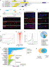
Figure 1.
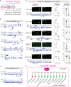
Figure 1.. Mapping genome-wide CFI-1 binding with ChIP-seq
(A) Schematic of CFI-1, Drosophila Dead ringer (Dri), and mouse Arid3a-c. The ARID (yellow) and REKLES (blue) domain are shown. (B) Diagram of the endogenous 3xflag::cfi-1 reporter allele. Bottom: dashed boxes highlight regions shown in (C)–(E). (C–E) Expression of the 3×FLAG::CFI-1 fusion protein is confirmed by immunostaining (DAPI, blue; anti-FLAG, red) in motor neurons (C), muscles (arrowheads) and head neurons (D), and tail neurons (E). Scale bars, 4 μm. (F) Fingerprint plot indicating localized strong enrichment of CFI-1 binding events in the genome. 50% of the maximum number of reads is contained in 86% of all genomic bins, indicating that 14% of the genome contains 50% of reads. (G) Heatmap of the CFI-1 ChIP-seq signal around 1.0 kb of the center of binding peaks. (H) Summary plot of the CFI-1 ChIP-seq signal; 95% confidence interval (gray area) around 3.0 kb of the TSS. Average signal peak is detected at ~140 bp upstream of the TSS. (I) Pie chart summarizing genomic distribution of the CFI-1 ChIP-seq signal. (J) Graph summarizing protein class ontology analysis (WormCat 2.0) of global CFI-1 target genes. 6,351 genes were analyzed, 4,678 of which have known protein class terms (1,673 genes were unassigned by WormCat 2.0). Asterisks to the left of each bar represent enriched categories (significantly overrepresented relative to the entire genome, Fisher’s exact test). Terminal differentiation genes, 2,947 of the 4,678 (63%); Gene expression, 806 of the 4,678 (17.2%). (K) Venn diagram showing that 77.1% of protein-coding genes (4,931 of 6,396) are bound by CFI-1 and expressed in cfi-1+ neurons based on RNA-seq (CenGEN data).
Figure 2.
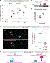
Figure 2.. CFI-1 directly activates terminal differentiation genes in IL2 sensory neurons
(A) Location of IL2 head sensory neurons. (B and C) IGV snapshots showing CFI-1 binding at loci of genes known to be activated (B) and repressed (C) by CFI-1 in IL2 neurons. (D) Expression analysis of five fluorescent markers of IL2 terminal differentiation (cil-7, cwp-4, ddn-3, degl-2, and tba-6) in WT and cfi-1 (ot786) mutant animals at L4. N = 10–15. Quantification of the number of IL2 neurons and fluorescence intensity is provided on the right. Scale bars, 4 μm. Two-tailed t test was performed. *p < 0.05, **p < 0.01, ***p < 0.001. CFI-1 binding at cil-7, cwp-4, ddn-3, degl-2, and tba-6 is shown below the microscopy images. (E) Model summarizing targets of CFI-1 in IL2 neurons.
Figure 3.
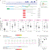
Figure 3.. CFI-1/Arid3a represses glr-4/GRIK4 in ventral cord motor neurons
(A) Summary of endogenous expression of cfi-1 and glr-4 in cholinergic motor neuron subtypes of the nerve cord (DA, DB, VA, VB, and AS) and retrovesicular ganglion (SAB). Expression patterns were determined by colocalization with neuron-subtype-specific reporters. See also Figure S6. Colored boxes represent expression; white boxes indicate no detectable expression. (B) CFI-1 binding signal on the glr-4 locus and design of the endogenous glr-4 reporter allele (2xNLS::mScarlet::SL2::glr-4). (C) Representative micrographs showing expression of 2×NLS:mScarlet:SL2:GLR-4 in SAB neurons of WT and cfi-1(−) animals. The 3 arrowheads point to the cell body of each SAB neuron. In cfi-1 (−) mutants, one SAB neuron is not located on the same focal plane with the other 2 SAB neurons. Hence, only 2 SAB neurons are shown. A bright white signal to the left of the SAB neurons indicates glr-4 expression in head neurons. (D) 2xNLS::mScarlet::SL2::glr-4 expression in ventral cord motor neurons (white arrowheads) in WT and cfi-1 (−) animals (L4). All MNs that express this reporter were counted. Ectopic expression of glr-4 was detected in cfi-1(−) mutants. Bottom: each dot in the graph of total number of MNs (left) represents an individual animal. Each dot in the fluorescence intensity quantification graph (right) represents an individual MN with glr-4 expression. Only MNs in the anterior nerve cord are included in the quantification. Two-tailed t test was performed. *p < 0.05, **p < 0.01, ***p < 0.001. N ≥ 15. (E) RNA-seq data (CeNGEN) showing expression of glr-4 in SAB, DB, VB, AS, VA, and DA motor neurons. (F) Single-molecule (sm) mRNA fluorescence in situ hybridization (FISH) for glr-4 in MNs of WT and cfi-1(−) animals (L1 stage). Left: images showing glr-4 mRNA (Cy5, red) in a motor neuron in cfi-1(−) mutants (nucleus: DAPI, blue). Right: quantification of the number of glr-4 transcripts in MNs of WT and cfi-1(−) animals. Two-tailed t test was performed. *p < 0.05, **p < 0.01, ***p < 0.001. Error bars, SEM. N ≥ 18. (G) RT-PCR for glr-4 in WT and cfi-1(−) mutants (whole-worm lysates). Error bars, SEM. (H) Model summarizing glr-4 regulation by CFI-1. Scale bars, 2 μm.
Figure 4.
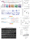
Figure 4.. CFI-1 is sufficient and continuously required to repress glr-4
(A) Quantification of the number of MNs expressing glr-4 in WT and cfi-1 (−) at four developmental stages: L2, L3, L4, and day 2 adults. Two-tailed t test was performed. *p < 0.05, **p < 0.01, ***p < 0.001. N ≥ 12. (B) Diagram showing the timeline of the auxin experiment. (C) Quantification of the endogenous glr-4 reporter in the ethanol group (control) and auxin group on kas16[mNG::AID::cfi-1]; otTi28[unc-11prom8+ehs-1prom7+rgef-1prom2::TIR1::mTurquoise2::unc-54 3′UTR] animals. otTi28 drives expression of TIR1 specifically in neurons. Two-tailed t test was performed. *p < 0.05, **p < 0.01, ***p < 0.001. N = 15. (D) Representative images showing loss of glr-4::tagRFP expression in SAB neurons (arrowheads) upon overexpression (OE) of cfi-1 with an SAB-specific promoter (unc-4). Right: quantification of the number of SAB neurons expressing glr-4::tagRFP. Two-tailed t test was performed. *p < 0.05, **p < 0.01, ***p < 0.001. N ≥ 14. Errorbars, SEM. Scale bar, 4 μm. (E) Model summarizing CFI-1 sufficiency in SAB neurons.
Figure 5.
Figure 5.. CFI-1 directly represses glr-4 by binding to its promoter via a conserved binding motif
(A) CFI-1 ChIP-seq tracks on the glr-4 locus. A series of glr-4 transgenic reporters is depicted. Right: the CFI-1 DNA binding motif. Using bioinformatics analysis, 13 CFI-1 binding motifs were identified upstream of glr-4 that overlap with CFI-1 binding peaks. (B) Quantification of MNs expressing glr-4 reporters in WT and cfi-1(−) animals. Two-tailed t test was performed. *p < 0.05, **p < 0.01, ***p < 0.001. All reporters were analyzed in the adult (day 2), except for the 538 bp and 4.9 kb reporters (analyzed at L4). N ≥ 13. Error bars, SEM. (C) Schematic of the 2xNLS::mScarlet::SL2::glr-411CFI–1 sites MUT allele. Point mutations were introduced to all 11 CFI-1 binding motifs at the glr-4 promoter. (D) Quantification of MNs expressing 2xNLS::mScarlet::SL2::glr-4 and 2xNLS::mScarlet::SL2::glr-411CFI–1 sites MUT alleles in day 2 adults. *p < 0.05, **p < 0.01, ***p < 0.001.
Figure 6.
Figure 6.. CFI-1 represses glr-4 by counteracting three terminal selectors
(A) Summary of the endogenous expression of cfi-1/Arid3, glr-4/GRIK4, unc-3/COE, lin-39 /HOX, and mab-5/HOX in five cholinergic motor neuron subtypes and SAB neurons. Colored boxes represent expression, while white boxes indicate no detectable expression. Only MNs located in the nerve cord are shown for DA, DB, VA, VB, and AS subtypes. (B) Quantification of 2xNLS::mScarlet::SL2::glr-4 expression in WT, cfi-1(−), lin-39(−); mab-5(−) double-mutant, and unc-3(−); lin-39(−); mab-5(−) triple-mutant animals (L4). Two-tailed t test. *p < 0.05, **p < 0.01, ***p < 0.001. N = 15. Error bars, SEM. (C) ChIP-seq binding peaks for UNC-3 (in WT and cfi-1(−)), CFI-1, LIN-39, and MAB-5 at the glr-4 locus. Point mutations were introduced to the proximal COE motif (UNC-3 binding site), resulting in the 2xNLS::mScarlet::SL2::glr-4COE1 MUT allele. (D) Quantification of the 2xNLS::mScarlet::SL2::glr-4 reporter in WT and unc-3(−) animals and of the 2xNLS::mScarlet::SL2::glr-4COE1 MUT reporter in day 2 adults. Two-tailed t test was performed. *p < 0.05, **p < 0.01, ***p < 0.001. N ≥ 13. (E) Quantification of the 2xNLS::mScarlet::SL2::glr-4 reporter in WT, cfi-1(−), unc-3(−), and cfi-1(−); unc-3(−) double mutants and of the 2xNLS::mScarlet::SL2::glr-4COE1 MUT allele in WT and cfi-1(−) animals (day 2 adults). Two-tailed t test was performed. *p < 0.05, **p < 0.01, ***p < 0.001. N ≥ 13. (F and G) Representative images (F) and fluorescence intensity quantification (G) of the 2xNLS::mScarlet::SL2::glr-4 reporter in WT, cfi-1(−), met-2(−), met-2(−); set-25(−), and cfi-1(−); met-2(−);set-25(−) animals at L4. Scale bars, 10 mm. In (F), a white dashed line indicates the C. elegans gut (autofluorescent). White arrowheads, MN nuclei expressing mScarlet. Two-tailed t test. *p < 0.05, **p < 0.01, ***p < 0.001. N ≥ 15.
Figure 7.
Figure 7.. Protein motif analysis of CFI-1
(A) Six CFI-1 constructs tested for rescue effects: WT cDNA, ΔARID, ΔeARID, ΔHTH, ΔARID + ΔeARID, and DREKLES. Two intrinsically disordered regions IDR1 (amino acids [aa] 4–56) and IDR2 (aa 121–157) were identified by NetSurfP-3.0 and PONDR (shown in gray). (B) Quantification of MNs showing expression of glr-4::tagrfp in cfi-1(ot786) mutant animals expressing the rescue constructs at L1. Two-tailed t test: *p < 0.05, **p < 0.01, ***p < 0.001. Error bars, SEM. (C) Schematic model summarizing our findings in SAB and nerve cord motor neurons.

Similar articles

Cited by

References

    1. Kortschak RD, Tucker PW, and Saint R (2000). ARID proteins come in from the desert. Trends Biochem. Sci 25, 294–299. 10.1016/s0968-0004(00)01597-8. - DOI - PubMed
    1. Patsialou A, Wilsker D, and Moran E (2005). DNA-binding properties of ARID family proteins. Nucleic Acids Res. 33, 66–80. 10.1093/nar/gki145. - DOI - PMC - PubMed
    1. Wilsker D, Patsialou A, Dallas PB, and Moran E (2002). ARID proteins: a diverse family of DNA binding proteins implicated in the control of cell growth, differentiation, and development. Cell Growth Differ. 13, 95–106. - PubMed
    1. Wilsker D, Probst L, Wain HM, Maltais L, Tucker PW, and Moran E (2005). Nomenclature of the ARID family of DNA-binding proteins. Genomics 86, 242–251. 10.1016/j.ygeno.2005.03.013. - DOI - PubMed
    1. Shang L, Cho MT, Retterer K, Folk L, Humberson J, Rohena L, Sidhu A, Saliganan S, Iglesias A, Vitazka P, et al. (2015). Mutations in ARID2 are associated with intellectual disabilities. Neurogenetics 16, 307–314. 10.1007/s10048-015-0454-0. - DOI - PubMed

Publication types

MeSH terms

Substances