. 2023 Mar 10;19(3):e1010843.
doi: 10.1371/journal.ppat.1010843. eCollection 2023 Mar.

Constitutive expression and distinct properties of IFN-epsilon protect the female reproductive tract from Zika virus infection

Affiliations

Constitutive expression and distinct properties of IFN-epsilon protect the female reproductive tract from Zika virus infection

Rosa C Coldbeck-Shackley et al. PLoS Pathog. .

Abstract

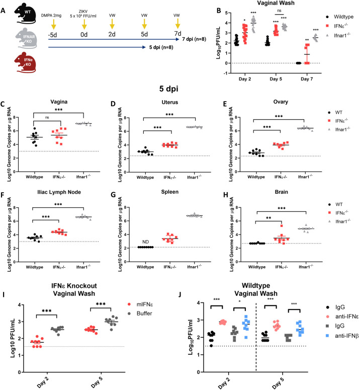
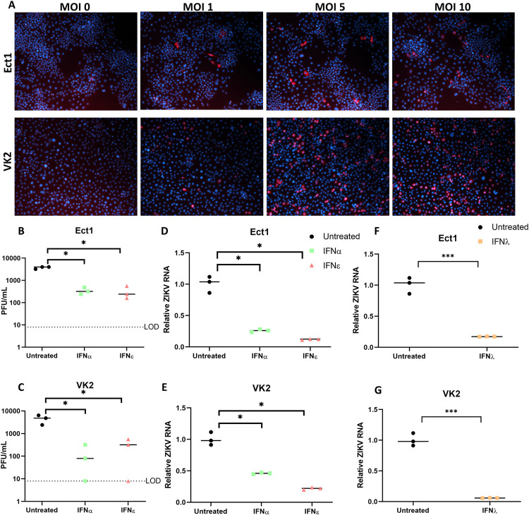
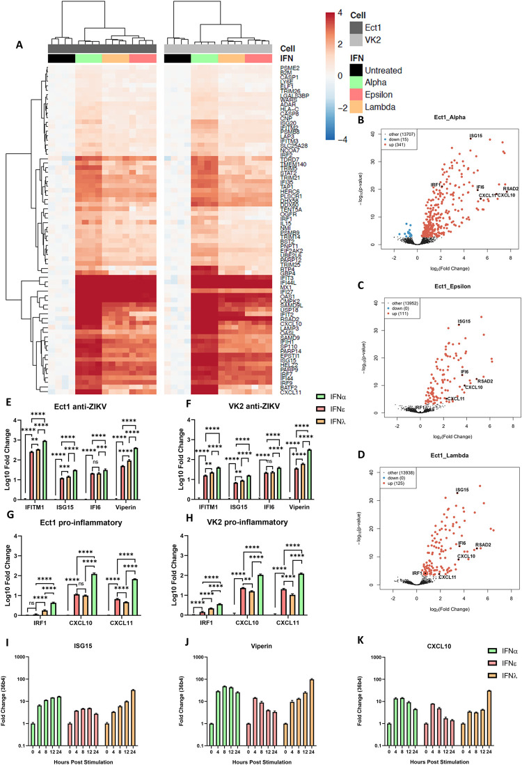
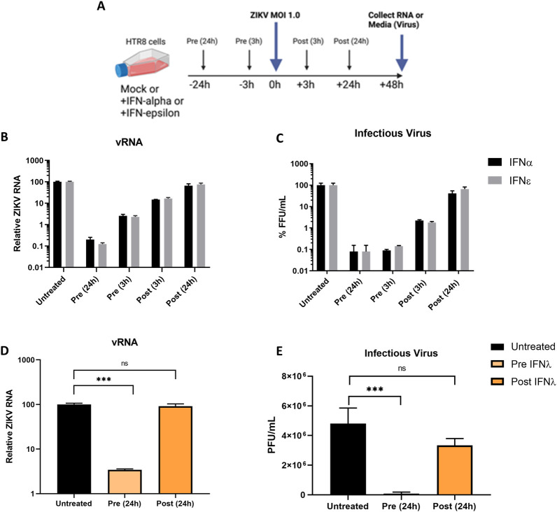
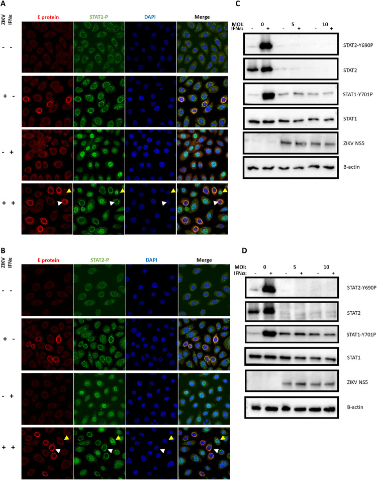
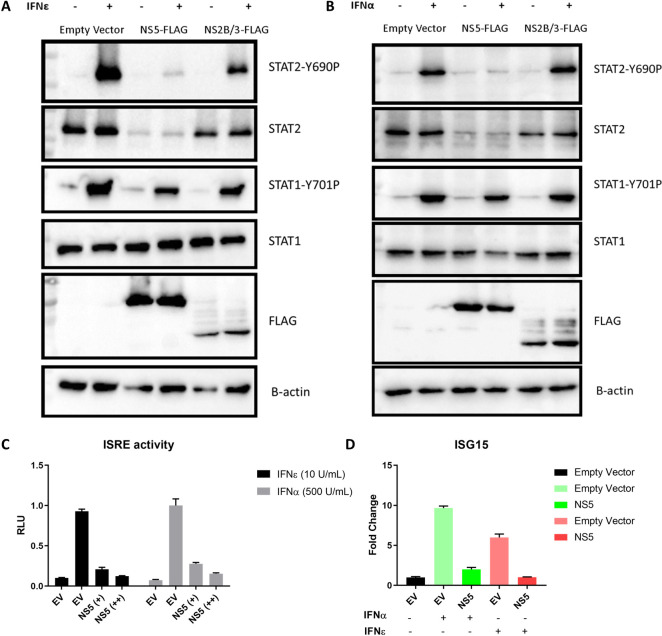
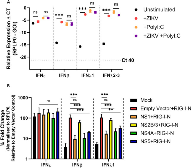
The immunological surveillance factors controlling vulnerability of the female reproductive tract (FRT) to sexually transmitted viral infections are not well understood. Interferon-epsilon (IFNɛ) is a distinct, immunoregulatory type-I IFN that is constitutively expressed by FRT epithelium and is not induced by pathogens like other antiviral IFNs α, β and λ. We show the necessity of IFNɛ for Zika Virus (ZIKV) protection by: increased susceptibility of IFNɛ-/- mice; their "rescue" by intravaginal recombinant IFNɛ treatment and blockade of protective endogenous IFNɛ by neutralising antibody. Complementary studies in human FRT cell lines showed IFNɛ had potent anti-ZIKV activity, associated with transcriptome responses similar to IFNλ but lacking the proinflammatory gene signature of IFNα. IFNɛ activated STAT1/2 pathways similar to IFNα and λ that were inhibited by ZIKV-encoded non-structural (NS) proteins, but not if IFNε exposure preceded infection. This scenario is provided by the constitutive expression of endogenous IFNε. However, the IFNɛ expression was not inhibited by ZIKV NS proteins despite their ability to antagonise the expression of IFNβ or λ. Thus, the constitutive expression of IFNɛ provides cellular resistance to viral strategies of antagonism and maximises the antiviral activity of the FRT. These results show that the unique spatiotemporal properties of IFNε provides an innate immune surveillance network in the FRT that is a significant barrier to viral infection with important implications for prevention and therapy.


Conflict of interest statement

The authors have declared that no competing interests exist.

Figures

Fig 1
Fig 1. ZIKV replication is inhibited by IFNε in a mouse model of vaginal transmission.
A) Experimental timeline of WT (black), mice lacking IFNε (IFNε-/-) (red) or mice lacking the type-I IFN receptor (IFNAR1-/-) (grey) were infected with ZIKV at 5 x 10⁵ FFU iVag 5 days post DMPA treatment and vaginal washes were taken at 2, 5, 7 dpi. Groups of 8 mice were culled at 5 dpi and 8 were culled at 7 dpi. B) Infectious virus was measured from vaginal washes by plaque assay at 2, 5, 7 dpi. C, D, E, F, G & H) Tissues taken at 5 dpi were used to harvest RNA for analysis of viral RNA by qRT-PCR in the vagina, uterus, ovary, illiac lymph node spleen and brain respectively. I) IFNε-/- mice were treated for 6h with either mIFNε or buffer prior to iVag infection with ZIKV, Infectious virus was measured from vaginal washes by plaque assay at 2 and 5 dpi. J) WT mice were treated for 6h with either an IFNε neutralising antibody (α-IFNε), an IFNβ (α-IFNβ) neutralising antibody, or a matched isotype control prior to iVag infection with ZIKV. Infectious virus was measured from vaginal washes by plaque assay at 2 and 5 dpi. Statistical analyses were performed on log10 transformed data using one-way ANOVA for multiple comparisons or t-test for two group comparisons. Data are presented as the mean +/- S.E.M. Schematic created with BioRender.com.
Fig 2
Fig 2. Ectocervical and Vaginal cell lines are permissive to ZIKV infection and treatment with IFNε is antiviral.
A) Ect1 or VK2 cells were infected with ZIKV PRVABC59 at the indicated MOI for 24h prior to staining, anti-flavi E staining (red), DAPI (blue). A-D) Ect1 or VK2 cells were treated overnight (16 hr) with 100 U/mL rhIFNε, rhIFNα-2A then infected with MOI 10 or 5 respectively for a further 48h prior to collecting supernatant and RNA for plaque assay (B & C) detection of infectious virus or qRT-PCR detection of vRNA (D & E). F-G) Ect1 or VK2 cells were treated overnight with 100 ng/mL rhFNλ-III then infected with MOI 10 or 5 respectively for a further 48h prior to harvesting RNA for detection of vRNA by qRT-PCR. Statistical analyses are performed by Brown-Forsythe and Welch ANOVA and multiple comparisons using a Dunnett’s T test compared to the untreated control (B-E) or by two tailed t-test (F & G), (n.s non-significant, * P < 0.05, ** P < 0.01). Data are presented as means with individual data points representative of independent biological replicates.
Fig 3
Fig 3. IFNε displays typical type-I IFN kinetics but induces an antiviral gene signature like IFNλ-3 at early time points in ectocervical and vaginal cells.
Ect1 and VK2 cells were treated with IFNε, IFNα-2a or IFNλ-3 (100 ng/mL) or left untreated (n = 4) for 6hr prior to RNA-seq analysis (NextSeq550 V2.5). Differentially expressed genes were determined with a 1.2-fold cut-off and adjusted p-value < 0.05. A) Heat map showing expression of key ISGs. B, C & D) Volcano plots indicating up or downregulation of genes after IFNα-2a, IFNε or IFNλ-3 treatment, respectively. E & F) Confirmation of anti-ZIKV ISGs (ISG15, IFI6, IFITM1, Viperin) expression by qRT-PCR. G & H) Confirmation of pro-inflammatory ISGs (IRF1, CXCL10, CXCL11) expression by qRT-PCR. Statistical analyses for E-G were performed separately for each gene by one-way ANOVA. I, J & K) Ect1 cells were treated with IFNε, IFNα-2a or IFNλ-3 (100 ng/mL) and RNA was harvested from untreated cells (t = 0) and at the indicated time points post stimulation. qRT-PCR was performed to detect expression of ISG15, Viperin and CXCL10.
Fig 4
Fig 4. ZIKV evades type-I and III IFN antiviral activity post-infection.
A) Timeline for IFN treatment regimes in HTR8 cells, arrows indicate media changes with either untreated media, IFNα, IFNε, or viral inoculum. B & C) HTR8 cells were infected with ZIKV PRVABC59 at a MOI of 1, cells were either primed with the indicated IFN (mIFNε 10 U/mL or hIFNα-2A 500 U/mL) or treated post infection. 48 hpi supernatants and RNA were harvested for determination of infectious virus by Focus Forming Assay or quantification of viral RNA by qRT-PCR. D & E) HTR8 cells were infected with ZIKV PRVABC59 at a MOI of 1, cells were either primed for 24h with hIFN λ-III (100 ng/mL) or treated post 24h post infection. Viral RNA and supernatant were collected 48hpi for determination of infectious virus by Focus Forming Assay or quantification of viral RNA by qRT-PCR. Statistical analyses are performed by or one-way ANOVA compared to the untreated control, (n.s non-significant, * P < 0.05, ** P < 0.01). Data are presented as means +/- S.D. Schematic created with BioRender.com.
Fig 5
Fig 5. Antiviral protection mediated by IFNε and other type-I IFNs is potently inhibited due to ZIKV inhibition of STAT1/2 signalling.
A & B) Ect1 cells were infected with ZIKV MOI of 10, 24 h post infection cells were stimulated with hIFNε (100 U/mL) for 30 min then fixed with acetone/methanol for detection of ZIKV E-antigen (red) and phosphorylated STAT1 or STAT2 proteins (green) by indirect immunofluorescence, DAPI (blue), infected cells (white arrows) and indicate uninfected bystanders (gold arrows). C & D) HeLa cells were infected at the indicated MOI of ZIKV 24h prior to stimulation with the either mIFNε (10 U/mL) or hIFNα-2A (500 U/mL) for 30 min, lysates were harvested for immunoblot of STAT1/2 protein and phosphorylated STAT1/2 proteins.
Fig 6
Fig 6. Evasion of IFNε antiviral activity is mediated by ZIKV NS5 degradation of STAT2.
A & B) HeLa cells were transfected with pCDNA6.2-ZIKV-NS5-FLAG, NS2B/3-FLAG or empty vector control and 24 h later stimulated for 30min with the indicated IFN prior to assessing total and phosphorylated STAT1/2 by immunoblotting. C) ISRE promoter activity was assessed following 24 h IFNε stimulation in the presence or absence of ZIKV NS5A expression. D) HeLa cells were transfected with pCDNA-NS5-FLAG expression plasmid or an empty vector control (EV), 24h post transfection cells were stimulated with the indicated type-I IFN for 6 h prior to harvesting RNA for qRT-PCR analysis of ISG15 expression.
Fig 7
Fig 7. IFNε RNA expression is not reduced by ZIKV infection or NS protein expression.
A) HeLa cells infected for 16h MOI 1 ZIKV PRV prior to stimulation with Poly I:C 1 μg for 8 h prior to assessing IFN gene expression by qRT-PCR. B) HeLa cells were co-transfected with expression plasmids encoding ZIKV NS1/2B/3/4A/5 or empty vector (pCDNA6.2) and the RIG-I-N plasmid or were mock transfected (Lipofectamine only), 24 h later RNA was harvested prior to assessing induction of type-I and type-III IFNs by qRT-PCR. Statistical analyses are performed by two-way ANOVA (A) or one-way ANOVA compared to the empty vector control (B), (n.s non-significant, * P < 0.05, ** P < 0.01). Data are presented as means +/- S.D.

References

    1. Sakkas H, Bozidis P, Giannakopoulos X, Sofikitis N, Papadopoulou C. An Update on Sexual Transmission of Zika Virus. Pathogens (Basel, Switzerland). 2018;7(3). Epub 2018/08/08. doi: 10.3390/pathogens7030066 ; PubMed Central PMCID: PMC6161238. - DOI - PMC - PubMed
    1. Pomar L, Lambert V, Madec Y, Vouga M, Pomar C, Matheus S, et al. Placental infection by Zika virus in French Guiana. Ultrasound in obstetrics & gynecology: the official journal of the International Society of Ultrasound in Obstetrics and Gynecology. 2019. Epub 2019/11/28. doi: 10.1002/uog.21936 . - DOI - PubMed
    1. Rasmussen SA, Jamieson DJ, Honein MA, Petersen LR. Zika Virus and Birth Defects—Reviewing the Evidence for Causality. The New England journal of medicine. 2016;374(20):1981–7. Epub 2016/04/14. doi: 10.1056/NEJMsr1604338 . - DOI - PubMed
    1. Russo FB, Jungmann P, Beltrao-Braga PCB. Zika infection and the development of neurological defects. Cellular microbiology. 2017;19(6). Epub 2017/04/04. doi: 10.1111/cmi.12744 . - DOI - PubMed
    1. Rice ME, Galang RR, Roth NM, Ellington SR, Moore CA, Valencia-Prado M, et al. Vital Signs: Zika-Associated Birth Defects and Neurodevelopmental Abnormalities Possibly Associated with Congenital Zika Virus Infection—U.S. Territories and Freely Associated States, 2018. MMWR Morbidity and mortality weekly report. 2018;67(31):858–67. Epub 2018/08/10. doi: 10.15585/mmwr.mm6731e1 ; PubMed Central PMCID: PMC6089332. - DOI - PMC - PubMed

Publication types