. 2023 Mar 14;120(11):e2207831120.
doi: 10.1073/pnas.2207831120. Epub 2023 Mar 10.

Propofol disrupts alpha dynamics in functionally distinct thalamocortical networks during loss of consciousness

Affiliations

Propofol disrupts alpha dynamics in functionally distinct thalamocortical networks during loss of consciousness

Veronica S Weiner et al. Proc Natl Acad Sci U S A. .

Abstract

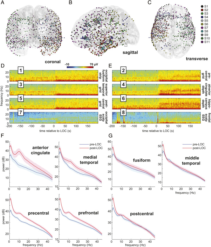
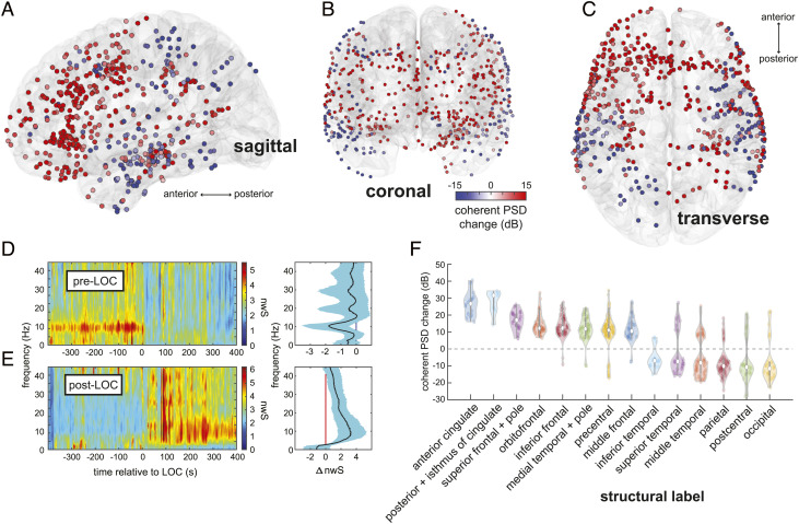
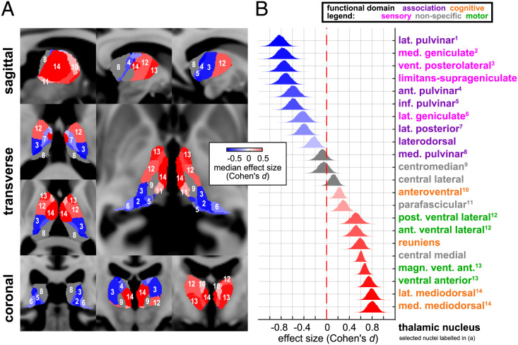
During propofol-induced general anesthesia, alpha rhythms measured using electroencephalography undergo a striking shift from posterior to anterior, termed anteriorization, where the ubiquitous waking alpha is lost and a frontal alpha emerges. The functional significance of alpha anteriorization and the precise brain regions contributing to the phenomenon are a mystery. While posterior alpha is thought to be generated by thalamocortical circuits connecting nuclei of the sensory thalamus with their cortical partners, the thalamic origins of the propofol-induced alpha remain poorly understood. Here, we used human intracranial recordings to identify regions in sensory cortices where propofol attenuates a coherent alpha network, distinct from those in the frontal cortex where it amplifies coherent alpha and beta activities. We then performed diffusion tractography between these identified regions and individual thalamic nuclei to show that the opposing dynamics of anteriorization occur within two distinct thalamocortical networks. We found that propofol disrupted a posterior alpha network structurally connected with nuclei in the sensory and sensory associational regions of the thalamus. At the same time, propofol induced a coherent alpha oscillation within prefrontal cortical areas that were connected with thalamic nuclei involved in cognition, such as the mediodorsal nucleus. The cortical and thalamic anatomy involved, as well as their known functional roles, suggests multiple means by which propofol dismantles sensory and cognitive processes to achieve loss of consciousness.

Keywords: alpha; intracranial EEG; propofol; synchrony; thalamocortical.


Conflict of interest statement

The authors have organizational affiliations to disclose, P.L.P. and E.N.B. are co-founders of PASCALL Systems, Inc., a start-up company developing closed-loop physiological control for anesthesiology. The authors have patent filings to disclose, P.L.P. and E.N.B. hold patents in general anesthesia monitoring technology.

Figures

Fig. 1.
Fig. 1.
Intracranial electrode coverage and spectral dynamics during propofol-induced general anesthesia. (AC) Intracranial recordings’ (n = 897) spatial coverage. Six intracranial channel locations from one subject are labeled. (D and E) Multitaper spectrograms in selected intracranial and scalp channels of a single subject (S7) aligned to LOC time. After LOC, a waking alpha rhythm in posterior regions (odd-numbered plots) dissipates, while a broader anesthesia-induced alpha rhythm emerges frontally (even-numbered plots). (F and G) Pre- and post-LOC multitaper spectra in selected frontal (F) and posterior (G) regions.
Fig. 2.
Fig. 2.
Spatiotemporal mapping of preanesthesia and postanesthesia coherent alpha networks. (AC) Changes in coherent alpha (10-Hz) cPSD across recording sites in all subjects. (D and E) Pre-LOC global coherence principal components depict a narrow 10-Hz rhythm disappearing at LOC (D), and a broader 10-Hz band beginning ~200 s after LOC (E). (F) cPSD is associated with structural (F) parcellations of brain regions. Frontal midline regions such as anterior and posterior cingulate, frontal, and orbitofrontal cortices, as well as the medial temporal lobe, show increased alpha-band cPSD after LOC, whereas posterior regions such as somatosensory and visual areas show a decrease. Constituent labels for each structural category are listed in SI Appendix, Table S3.
Fig. 3.
Fig. 3.
Distinct thalamocortical connections underlying preanesthesia and postanesthesia alpha networks. (A) Thalamic nuclei are selectively connected to distinct networks showing either increased or decreased cPSD after LOC. (B) Primary and association sensory thalamic nuclei selectively connect to alpha cPSD-decreasing regions; nuclei of the executive and cognitive thalamus selectively connect to alpha cPSD-increasing regions.

References

    1. Feshchenko V. A., Veselis R. A., Reinsel R. A., Propofol-induced alpha rhythm. Neuropsychobiology 50, 257–266 (2004). - PubMed
    1. Ching S., Cimenser A., Purdon P. L., Brown E. N., Kopell N. J., Thalamocortical model for a propofol-induced α-rhythm associated with loss of consciousness. Proc. Natl. Acad. Sci. U.S.A. 107, 22665–22670 (2010). - PMC - PubMed
    1. Cimenser A., et al. , Tracking brain states under general anesthesia by using global coherence analysis. Proc. Natl. Acad. Sci. U.S.A. 108, 8832–8837 (2011). - PMC - PubMed
    1. Purdon P. L., et al. , Electroencephalogram signatures of loss and recovery of consciousness from propofol. Proc. Natl. Acad. Sci. U.S.A. 110, E1142–E1151 (2013). - PMC - PubMed
    1. Flores F. J., et al. , Thalamocortical synchronization during induction and emergence from propofol-induced unconsciousness. Proc. Natl. Acad. Sci. U.S.A. 114, E6660–E6668 (2017). - PMC - PubMed

Publication types

LinkOut - more resources