. 2023 Apr 20;30(4):394-402.e9.
doi: 10.1016/j.chembiol.2023.02.008. Epub 2023 Mar 9.

Chemoproteomics-enabled discovery of a covalent molecular glue degrader targeting NF-κB

Affiliations

Chemoproteomics-enabled discovery of a covalent molecular glue degrader targeting NF-κB

Elizabeth A King et al. Cell Chem Biol. .

Abstract

Targeted protein degradation has arisen as a powerful therapeutic modality for degrading disease targets. While proteolysis-targeting chimera (PROTAC) design is more modular, the discovery of molecular glue degraders has been more challenging. Here, we have coupled the phenotypic screening of a covalent ligand library with chemoproteomic approaches to rapidly discover a covalent molecular glue degrader and associated mechanisms. We have identified a cysteine-reactive covalent ligand EN450 that impairs leukemia cell viability in a NEDDylation and proteasome-dependent manner. Chemoproteomic profiling revealed covalent interaction of EN450 with an allosteric C111 in the E2 ubiquitin-conjugating enzyme UBE2D. Quantitative proteomic profiling revealed the degradation of the oncogenic transcription factor NFKB1 as a putative degradation target. Our study thus puts forth the discovery of a covalent molecular glue degrader that uniquely induced the proximity of an E2 with a transcription factor to induce its degradation in cancer cells.

Keywords: E2 ligase; NFKB1; UBE2D; activity-based protein profiling; molecular glue; targeted protein degradation; transcription factor.


Conflict of interest statement

Declaration of interests J.A.T., J.M.K., and D.D. are employees of Novartis Institutes for BioMedical Research. This study was funded by the Novartis Institutes for BioMedical Research and the Novartis-Berkeley Translational Chemical Biology Institute. D.K.N. is a co-founder, shareholder, and on the scientific advisory boards for Frontier Medicines and Vicinitas Therapeutics; a member of the board of directors for Vicinitas Therapeutics; on the scientific advisory boards of The Mark Foundation for Cancer Research, Photys Therapeutics, Apertor Pharmaceuticals, Ecto Therapeutics, Oerth Bio, and Chordia Therapeutics; and on the investment advisory board of Droia Ventures.

Figures

Figure 1.
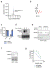
Figure 1.. Discovery of a covalent molecular glue degrader with anti-proliferative activities in HAP1 leukemia cancer cells.
a) HAP1 cell viability screen of cysteine-reactive covalent ligands. DMSO vehicle or cysteine-reactive covalent ligands (50 μM) were treated in HAP1 cells for 24 h and cell viability was assessed by Hoescht staining. Compounds highlighted in red and labeled are those that impaired HAP1 cell viability by greater than 90 % compared to vehicle-treated controls. This screen was performed with n=1 biological replicate/group. (b) The 11 hit compounds from (a) were rescreened with n=3 biologically independent replicates/group in HAP1 cells under the same conditions. Individual replicate values shown. Among these hits EN222, EN226, and EN450 showed reproducible impaired HAP1 cell viability of greater than 90 % compared to DMSO vehicle-treated controls. (c) Knockdown of UBE2M. HAP1 cells were transiently transfected with siControl or siUBE2M oligonucleotides and knockdown of UBE2M was assessed by Western blotting compared to GAPDH loading control. (d) HAP1 cell viability in siControl and siUBE2M cells treated with DMSO vehicle, the positive control molecular glue degrader dCEMM1, EN222, EN266, or EN450 (50 μM) for 24 h, assessed by Hoescht staining. (e) structure of EN450 with the reactive acrylamide handle highlighted in red. (f) Attenuation of HAP1 cell viability impairments by NEDDylation inhibitor MLN4924. HAP1 cells were pre-treated with DMSO vehicle or MLN4924 (1 μM) for 1 h prior to treatment of cells with DMSO vehicle, dCEMM1, or EN450 (50 μM) for 24 h, and cell viability was assessed by Hoechst staining. (g) Attenuation of HAP1 cell viability impairments by proteasome inhibitor bortezomib. HAP1 cells were pre-treated with DMSO vehicle or bortezomib (1 μM) for 1 h prior to treatment of cells with DMSO vehicle or EN450 (50 μM) for 24 h, and cell viability was assessed by Hoechst staining. Data in (d, f, g) are average ± sem of n=3-6 biologically independent replicates/group with individual replicate values also shown. Statistical significance is expressed as *p<0.05 compared to siControl or DMSO control groups in (d, f, g), #p<0.05 compared to corresponding siControl or DMSO pre-treated treatment groups, and calculated with Student’s unpaired two-tailed t-tests. Related to Table S1.
Figure 2.
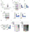
Figure 2.. Chemoproteomic profiling and validation of EN450 targets in HAP1 cells.
(a) Cysteine chemoproteomic profiling of EN450 targets in HAP1 cells using isoTOP-ABPP. HAP1 cells were treated with DMSO vehicle or EN450 (50 μM) for 3 h, after which resulting cell lysates were labeled with an alkyne-functionalized iodoacetamide cysteine-reactive probe (200 μM) for 1 h, and an isotopically light (for DMSO) or heavy (for EN450) biotin-azide handle bearing a TEV protease recognition peptide was appended by CuAAC. Control and treated proteomes were combined in a 1:1 ratio, taken through the isoTOP-ABPP procedure and light/heavy probe-modified peptides were analyzed by LC-MS/MS and quantified. Shown in blue are probe-modified peptides with light/heavy ratios >1.3 with adjusted p<0.05. Shown in red is C111 of UBE2D. Data shown are average ratios from n=4 biologically independent replicates/group. (b) structure of alkyne-functionalized probe derivative of EN450—EK-1-8 with cysteine-reactive acrylamide warhead highlighted in red. (c) Attenuation of HAP1 cell viability impairments by NEDDylation inhibitor MLN4924. HAP1 cells were pre-treated with DMSO vehicle or MLN4924 (1 μM) for 1 h prior to treatment of cells with DMSO vehicle, EK-1-8 (100 μM) for 24 h, and cell viability was assessed by Hoechst staining. (d) EK-1-8 labeling of recombinant pure human UBE2D1 C85S mutant protein. UBE2D1 C85S mutant protein was labeled with DMSO vehicle or EK-1-8 for 30 min, after which an azide functionalized rhodamine handle was appended onto probe-labeled protein by CuAAC, after which proteins were resolved by SDS/PAGE and visualized by in-gel fluorescence. Protein loading was assessed by silver staining. Gels are representative of an n=3 biologically independent replicates/group. (e) UBE2D1 pulldown from HAP1 cells with EK-1-8 probe. HAP1 cells were pre-treated with DMSO vehicle or EN450 (50 μM) for 1 h prior to treatment of cells with DMSO vehicle or EK-1-8 (10 μM) for 3 h. Probe-labeled proteins from resulting cell lysates were subsequently appended to an azide-functionalized biotin handle by CuAAC, avidin-enriched, eluted and separated by SDS/PAGE and blotted for UBE2D1 and unrelated protein GAPDH. Shown on the blot are also input UBE2D1 and GAPDH protein levels between the three groups shown. Blots are representative of an n=3 biologically independent replicates/group. (f) Confirmation of UBE2D1 knockdown. HAP1 cells were transiently transfected with siControl or siUBE2D1 oligonucleotides and UBE2D1 and loading control GAPDH expression were assessed after 72 h by Western blotting. Blot is representative of n=3 biologically independent replicates/group. (g) Percent HAP1 cell proliferation in siControl and siUBE2D1 cells treated with EN450 for 24 h compared to DMSO vehicle-treated controls. Data shown in (c) and (g) are average ± sem of n=6-30 biologically independent replicates/group with individual replicate values also shown. Statistical significance in (c) is expressed as *p<0.05 compared to DMSO-pre-treated control groups and #p<0.05 compared to DMSO-pre-treated EK-1-8-treated groups and in (g) is expressed as *p<0.05 compared to the corresponding treatment group in siControl cells. Related to Table S2, Figure S1, and Figure S2.
Figure 3.
Figure 3.. Identification of the protein degraded by EN450.
(a) TMT-based quantitative proteomic profiling of EN450 in HAP1 cells. HAP1 cells were treated with DMSO vehicle or EN450 (50 μM) for 24 h. Shown in red is the only protein reduced in levels by >4-fold with p-value <0.05—NFKB1. Data shown are from n=3 biologically independent replicates/group. (b) Western blotting analysis of p105 and p50 isoforms of NFKB1 and loading control vinculin levels in HAP1 cells treated with DMSO vehicle or EN450 (50 μM) for 24 h. (c, d) Quantification of experiment described in (b). (e) NEDDylation inhibitor attenuates NFKB1 loss by EN450 treatment in HAP1 cells. HAP1 cells were pre-treated with DMSO vehicle or MLN4924 (1 μM) for 1 h prior to treatment of cells with DMSO vehicle or EN450 (50 μM) for 24 h. NFKB1 and loading control vinculin levels were assessed by Western blotting. (f) Quantification of experiment described in (e). (g) Ternary complex formation between UBE2D1, EN450, and NFKB1. Recombinant pure human GST-NFKB1 and UBE2D1 proteins were incubated with DMSO vehicle or EN450 (50 μM) for 1 h, after which GST-NFKB1 was enriched and the pulldown eluate was blotted for NFKB1 and UBE2D1. (h) Quantification of pulldown experiment described in (g). (i, j) Ubiquitination assay with UBE2D1, ATP, FLAG-Ubiquitin, and NFKB1 pure proteins with the exclusion or addition of the CUL4A/RBX1/NEDD8 Cullin complex. This incubation was treated with DMSO vehicle or EN450 (50 μM) for 1 h. NFKB1 ubiquitination was detected by Western blotting visualizing FLAG-Ub in (i) and NFKB1 in (j). Blots shown in (b, e, g) are representative of n=3 biologically independent replicates/group. Data shown in (c, d, f, h) are average ± sem of n=3-8 biologically independent replicates/group with individual replicate values also shown. Statistical significance in (c, d, f, h) are expressed as *p<0.05 compared to DMSO vehicle-treated controls and NS denotes not significant. Related to Table S3 and Figure S3.
Figure 4.
Figure 4.. Understanding the role of NFKB1 in EN450-mediated effects.
(a) Stable lentiviral overexpression of FLAG-NFKB1 in HAP1 cells assessed by Western blotting for NFKB1, FLAG, and loading control GAPDH. (b) HAP1 eGFP control or FLAG-NFKB1 overexpressing cell viability from cells treated with DMSO vehicle or EN450 for 24 h. (c) Attenuation of HEK293T cell viability impairments by NEDDylation inhibitor MLN4924. HEK293T cells were pre-treated with DMSO vehicle or MLN4924 (1 μM) for 1 h prior to treatment of cells with DMSO vehicle or EN450 (50 μM) for 24 h, and cell viability was assessed by Hoechst staining. (d) NFKB1 and loading control vinculin levels in HEK293T cells pre-treated with DMSO vehicle or MLN4924 (1 μM) 1 h prior to treatment of cells with DMSO vehicle or EN450 (50 μM) for 24 h, assessed by Western blotting. (e) Transient transfection and overexpression of FLAG-NFKB1 in HEK293T cells assessed by Western blotting of NFKB1, FLAG, and loading control GAPDH. (f) HEK293T eGFP control or FLAG-NFKB1 overexpressing cell viability from cells treated with DMSO vehicle or EN450 (50 μM) for 24 h. Blots shown in (a, d, e) are representative of n=3 biologically independent replicates/group. Data shown in (c, f) are average ± sem of n=3-8 biologically independent replicates/group with individual replicate values also shown. Statistical significance in (c, f) are expressed as *p<0.05 compared to DMSO vehicle-treated controls or eGFP-expressing control cells and as #p<0.05 compared to DMSO pre-treated EN450-treated groups in (c) or EN450-treated eGFP-expressing cells in (f).

Comment in

Similar articles

Cited by

References

    1. Spradlin JN, Zhang E, and Nomura DK (2021). Reimagining Druggability Using Chemoproteomic Platforms. Acc. Chem. Res 54, 1801–1813. 10.1021/acs.accounts.1c00065. - DOI - PubMed
    1. Dixon SJ, and Stockwell BR (2009). Identifying druggable disease-modifying gene products. Curr. Opin. Chem. Biol 13, 549–555. 10.1016/j.cbpa.2009.08.003. - DOI - PMC - PubMed
    1. Bond MJ, and Crews CM (2021). Proteolysis targeting chimeras (PROTACs) come of age: entering the third decade of targeted protein degradation. RSC Chem. Biol 2, 725–742. 10.1039/d1cb00011j. - DOI - PMC - PubMed
    1. Burslem GM, and Crews CM (2020). Proteolysis-Targeting Chimeras as Therapeutics and Tools for Biological Discovery. Cell 181, 102–114. 10.1016/j.cell.2019.11.031. - DOI - PMC - PubMed
    1. Schreiber SL (2021). The Rise of Molecular Glues. Cell 184, 3–9. 10.1016/j.cell.2020.12.020. - DOI - PubMed

Publication types