. 2023 Mar 10;10(1):129.
doi: 10.1038/s41597-023-02002-8.

Neonatal EEG graded for severity of background abnormalities in hypoxic-ischaemic encephalopathy

Affiliations

Neonatal EEG graded for severity of background abnormalities in hypoxic-ischaemic encephalopathy

John M O'Toole et al. Sci Data. .

Abstract

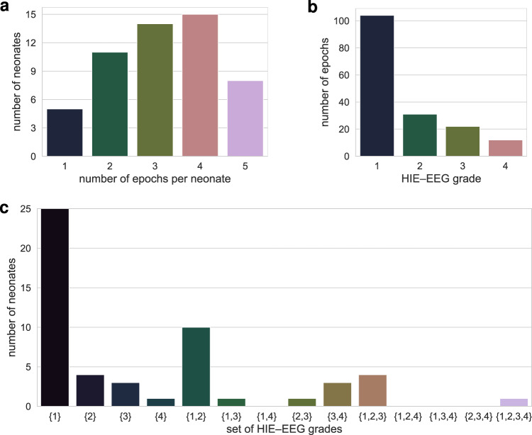
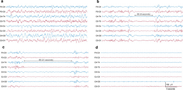
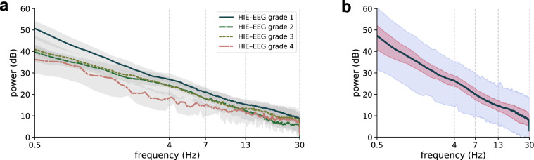
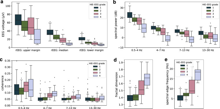
This report describes a set of neonatal electroencephalogram (EEG) recordings graded according to the severity of abnormalities in the background pattern. The dataset consists of 169 hours of multichannel EEG from 53 neonates recorded in a neonatal intensive care unit. All neonates received a diagnosis of hypoxic-ischaemic encephalopathy (HIE), the most common cause of brain injury in full term infants. For each neonate, multiple 1-hour epochs of good quality EEG were selected and then graded for background abnormalities. The grading system assesses EEG attributes such as amplitude, continuity, sleep-wake cycling, symmetry and synchrony, and abnormal waveforms. Background severity was then categorised into 4 grades: normal or mildly abnormal EEG, moderately abnormal EEG, majorly abnormal EEG, and inactive EEG. The data can be used as a reference set of multi-channel EEG for neonates with HIE, for EEG training purposes, or for developing and evaluating automated grading algorithms.


Conflict of interest statement

The authors declare no competing interests.

Figures

Fig. 1
Fig. 1
Distribution of EEG epochs. One-hundred and sixty nine  1-hour epochs were pruned from continuous EEG recordings from 53 neonates. Distribution of epochs per baby in (a) and grades of EEG hypoxic-ischaemic encephalopathy (HIE) in (b). Some neonates have more than one grade throughout the epochs: (c) illustrates the distribution of all possible combinations of sets of grades per neonate.
Fig. 2
Fig. 2
Examples of different EEG grades. Thirty-six seconds of EEG from different neonates. (a) normal or mildly abnormal EEG (grade 1); (b) moderately abnormal EEG (grade 2); (c) major abnormalities (grade 3); (d) inactive EEG (grade 4). Inter-burst intervals (IBI) are annotated in the grade 2 and grade 3 examples. All EEGs are in bipolar montage, plotted with the same time and amplitude scale, and bandpass filtered from 0.3 to 35 Hz.
Fig. 3
Fig. 3
Examples of some typical EEG artefacts. Thirty-six seconds of EEG from different neonates. EEG segment with (a) high-amplitude movement artefact (grade 1 EEG); (b) sweat artefact (also grade 1 EEG);(c) ECG artefact on C4–O2 and C4–T4 and high-frequency muscle artefact, most prominent on F3–C3 (grade 3 EEG); (d) respiration artefact across the left hemisphere channels, clearly visible on C3–O1 (grade 4 EEG). All EEGs are in bipolar montage and bandpass filtered from 0.1 to 35 Hz.
Fig. 4
Fig. 4
Spectra plotted on a log–log scale from epochs for each grade (a) and a grand-average for all epochs (b). Thick lines represent the median value across all epochs, and shaded areas represent the inter-quartile range in (a) and (b) and 95-th centile range in (b). There are 104 epochs for grade 1, 31 for grade 2, 22 for grade 3, and 12 for grade 4.
Fig. 5
Fig. 5
Quantitative summary measures of the EEG for the 4 HIE–EEG grades. Three summary measures of the range-EEG (rEEG) in (a), spectral power in (b), inter-hemispheric coherence in (c), fractal dimension in (d), and spectral edge frequency in (e). Spectral power and coherence features are calculated for 4 different frequency bands.

References

    1. Kurinczuk JJ, White-Koning M, Badawi N. Epidemiology of neonatal encephalopathy and hypoxic-ischaemic encephalopathy. Early Hum. Dev. 2010;86:329–338. doi: 10.1016/j.earlhumdev.2010.05.010. - DOI - PubMed
    1. Perez A, et al. Long-term neurodevelopmental outcome with hypoxic-ischemic encephalopathy. J. Pediatr. 2013;163:454–459. doi: 10.1016/j.jpeds.2013.02.003. - DOI - PubMed
    1. Walsh BH, Murray DM, Boylan GB. The use of conventional EEG for the assessment of hypoxic ischaemic encephalopathy in the newborn: a review. Clin. Neurophysiol. 2011;122:1284–1294. doi: 10.1016/j.clinph.2011.03.032. - DOI - PubMed
    1. Holmes G, et al. Prognostic value of the electroencephalogram in neonatal asphyxia. Electroencephalogr. Clin. Neurophysiol. 1982;53:60–72. doi: 10.1016/0013-4694(82)90106-7. - DOI - PubMed
    1. Pressler RM, Boylan GB, Morton M, Binnie CD, Rennie JM. Early serial EEG in hypoxic ischaemic encephalopathy. Clin. Neurophysiol. 2001;112:31–37. doi: 10.1016/S1388-2457(00)00517-4. - DOI - PubMed