. 2023 Mar 1;13(5):891.
doi: 10.3390/ani13050891.

Hepatitis E Virus (HEV) Spreads from Pigs and Sheep in Mongolia

Affiliations

Hepatitis E Virus (HEV) Spreads from Pigs and Sheep in Mongolia

Enkhbaatar Batmagnai et al. Animals (Basel). .

Abstract

Hepatitis E is a viral infectious disease in pigs, wild boars, cows, deer, rabbits, camels, and humans as hosts caused by Paslahepevirus. Recently, it has been detected in a wide variety of animals including domestic small ruminants. Mongolia is a land of nomadic people living with livestock such as sheep, goats, and cattle. Due to how Mongolian lifestyles have changed, pork has become popular and swine diseases have emerged. Among them, Hepatitis E disease has become a zoonotic infectious disease that needs to be addressed. The HEV problem in pigs is that infected pigs excrete the virus without showing clinical symptoms and it spreads into the environment. We attempted to detect HEV RNA in sheep which had been raised in Mongolia for a long time, and those animals living together with pigs in the same region currently. We also conducted a longitudinal analysis of HEV infection in pigs in the same area and found that they were infected with HEV of the same genotype and cluster. In this study, we examined 400 feces and 120 livers (pigs and sheep) by RT-PCR in Töv Province, Mongolia. HEV detection in fecal samples was 2% (4/200) in sheep and 15% (30/200) in pigs. The results of ORF2 sequence analysis of the HEV RT-PCR-positive pigs and sheep confirmed genotype 4 in both animals. The results suggest that HEV infection is widespread in both pigs and sheep and that urgent measures to prevent infection are needed. This case study points to the changing nature of infectious diseases associated with livestock farming. It will be necessary to reconsider livestock husbandry and public health issues based on these cases.

Keywords: Mongolia; hepatitis E; phylogenetic analysis; pig; prevalence; sheep.


Conflict of interest statement

The authors declare no conflict of interest.

Figures

Figure 1
Figure 1
Sampling locations in Mongolia. The upper left figure shows the whole Mongolian territory with two neighboring countries and the locations of Töv Province and the capital city, Ulaanbaatar. The lower left figure shows the whole territory of Ulaanbaatar city and the location of the pig farm in Songino Khairkhan district in the red rectangle. The figure on the right shows the whole territory of Töv Province and the locations of four soums are shown in pink (Zaamar, Undurshireet, Bayanchandmani, and Batsumber soums) where we collected the feces and liver samples of the sheep. In addition, the figure shows that the Bayanchandmani and Batsumber soums are near to the pig farm located in Songino Khairkhan district. Emeelt—the main slaughterhouse in Mongolia—is also in Songino Khairkhan district. The red rectangles show the areas where the pig feces samples were collected.
Figure 2
Figure 2
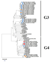
Phylogenetic analysis of HEV-ORF2 regions detected in animals in Mongolia with Smith’s prototype sequences. Phylogenetic tree (neighbor-joining method) of the open reading frame (ORF) 2 gene region of hepatitis E virus (HEV) genotypes 3 and 4. The numbers and letters before the accession number in the table indicate the HEV subtype, and the genotype (HEV-3, HEV-4) is shown after the sample number identified in this study. Blue dots represent pig feces samples, orange dots represent pig liver samples, and red dots represent sheep liver samples (ShL-0-2). A total of 28 samples were sequenced for HEV ORF2. MEGA X was used for multiple alignments and phylogenetic tree construction. Based on the phylogenetic tree results, 9/28 sequences belonged to HEV-4, whereas 19/28 sequences belonged to HEV-3. The ORF2 gene region of the JQ001749.1_Bat HEV/GE/2009 (GE—Germany) complete genome was used as an outgroup. The bar represents 0.05 nucleotide substitutions per site.
Figure 3
Figure 3
The Bayesian maximum clade credibility (MCC) host discrete traits tree for 302 bp HEV ORF2 nucleotide sequences (nt position 6022–6324) of 40 Mongolian strains, including 28 sequences from our study and reference strains (six swine HEV-ORF2 genotype 3, reprinted with permission from reference [16], four human HEV-ORF2 sequences [19], and two Bactrian camels HEV-ORF2 (BcHEV1, BcHEV2) sequences [26]) obtained from the GenBank database. The phylogenetic host tree indicated transmission between HEV hosts, the host at the node indicated the ancestor of the sub-group, and the number at the node indicated the posterior probability. The bar at the bottom of the figure denotes evolutionary distance.

References

    1. Purdy M.A., Drexler J.F., Meng X.J., Norder H., Okamoto H., Van der Poel W.H.M., Reuter G., de Souza W.M., Ulrich R.G., Smith D.B. ICTV Virus Taxonomy Profile: Hepeviridae 2022. J. Gen. Virol. 2022;103:1–2. doi: 10.1099/jgv.0.001778. - DOI - PubMed
    1. Li S., Liu M., Cong J., Zhou Y., Miao Z. Detection and Characterization of Hepatitis e Virus in Goats at Slaughterhouse in Tai’an Region, China. BioMed Res. Int. 2017;2017:3723650. doi: 10.1155/2017/3723650. - DOI - PMC - PubMed
    1. Hu G.D., Ma X. Detection and Sequences Analysis of Bovine Hepatitis E Virus RNA in Xinjiang Autonomous Region. Bing Du Xue Bao. 2010;26:27–32. - PubMed
    1. Huang F., Li Y., Yu W., Jing S., Wang J., Long F., He Z., Yang C., Bi Y., Cao W., et al. Excretion of Infectious Hepatitis E Virus into Milk in Cows Imposes High Risks of Zoonosis. Hepatology. 2016;64:350–359. doi: 10.1002/hep.28668. - DOI - PubMed
    1. Wu J., Si F., Jiang C., Li T., Jin M. Molecular Detection of Hepatitis E Virus in Sheep from Southern Xinjiang, China. Virus Genes. 2015;50:410–417. doi: 10.1007/s11262-015-1194-9. - DOI - PMC - PubMed

LinkOut - more resources