An official website of the United States government
The .gov means it’s official.
Federal government websites often end in .gov or .mil. Before
sharing sensitive information, make sure you’re on a federal
government site.
The site is secure.
The https:// ensures that you are connecting to the
official website and that any information you provide is encrypted
and transmitted securely.
Hypertrophic scarring (HTS) is an aberrant form of wound healing that is associated with excessive deposition of extracellular matrix and connective tissue at the site of injury. In this review article, we provide an overview of normal (acute) wound healing phases (hemostasis, inflammation, proliferation, and remodeling). We next discuss the dysregulated and/or impaired mechanisms in wound healing phases that are associated with HTS development. We next discuss the animal models of HTS and their limitations, and review the current and emerging treatments of HTS.
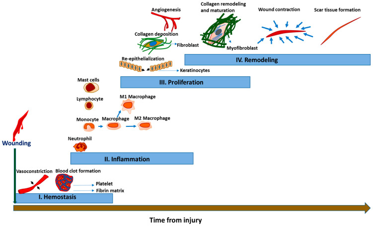
The phases of acute wound healing, including hemostasis (I), inflammation (II), proliferation (III),…
Figure 1
The phases of acute wound healing, including hemostasis (I), inflammation (II), proliferation (III), and remodeling (IV). Hemostasis begins soon after wounding with vasoconstriction and blood clot formation. This is followed by the infiltration of inflammatory cells. Then, re-epithelialization occurs with collagen deposition and angiogenesis during the proliferation phase. Finally, the remodeling phase occurs with collagen remodeling and maturation, wound contraction, and scar tissue formation.
Figure 2
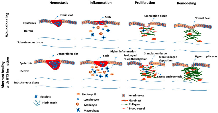
Phases of normal wound healing…
Figure 2
Phases of normal wound healing versus aberrant wound healing with the formation of…
Figure 2
Phases of normal wound healing versus aberrant wound healing with the formation of hypertrophic scars. Events such as higher fibrin clot deposition, infiltration of higher number of inflammatory cells, prolonged re-epithelialization, and excess angiogenesis can result in excessive and improper collagen deposition and, therefore, formation of hypertrophic scars.
Figure 3
The events and molecules associated…
Figure 3
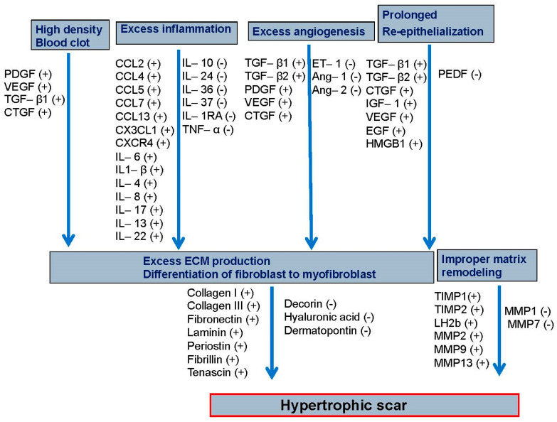
The events and molecules associated with hypertrophic scar (HTS) formation. The pro-fibrotic molecules…
Figure 3
The events and molecules associated with hypertrophic scar (HTS) formation. The pro-fibrotic molecules generated from the high-density blood clot induce excessive inflammation, angiogenesis, and prolonged re-epithelization. The resultant excessive production of extracellular matrix and fibroblast differentiation and improper matrix remodeling then causes formation of hypertrophic scarring. Molecules with increased expression are denoted with (+), whereas those with decreased expression are denoted with (−). The players included in this figure are discussed in the text.
Gurtner G.C., Werner S., Barrandon Y., Longaker M.T. Wound repair and regeneration. Nature. 2008;453:314–321. doi: 10.1038/nature07039.
-
DOI
-
PubMed
Diegelmann R.F., Evans M.C. Wound healing: An overview of acute, fibrotic and delayed healing. Front. Biosci. 2004;9:283–289. doi: 10.2741/1184.
-
DOI
-
PubMed
Ogawa R., Akita S., Akaishi S., Aramaki-Hattori N., Dohi T., Hayashi T., Kishi K., Kono T., Matsumura H., Muneuchi G. Diagnosis and treatment of keloids and hypertrophic scars—Japan scar workshop consensus document 2018. Burn. Trauma. 2019;7:39. doi: 10.1186/s41038-019-0175-y.
-
DOI
-
PMC
-
PubMed
Limandjaja G.C., Niessen F.B., Scheper R.J., Gibbs S. Hypertrophic scars and keloids: Overview of the evidence and practical guide for differentiating between these abnormal scars. Exp. Dermatol. 2021;30:146–161. doi: 10.1111/exd.14121.
-
DOI
-
PMC
-
PubMed
Marshall C.D., Hu M.S., Leavitt T., Barnes L.A., Lorenz H.P., Longaker M.T. Cutaneous scarring: Basic science, current treatments, and future directions. Adv. Wound Care. 2018;7:29–45. doi: 10.1089/wound.2016.0696.
-
DOI
-
PMC
-
PubMed