Review
. 2023 Feb 21;12(5):682.
doi: 10.3390/cells12050682.

A Beginner's Guide to Cell Culture: Practical Advice for Preventing Needless Problems

Affiliations
Review

A Beginner's Guide to Cell Culture: Practical Advice for Preventing Needless Problems

Sabine Weiskirchen et al. Cells. .

Abstract

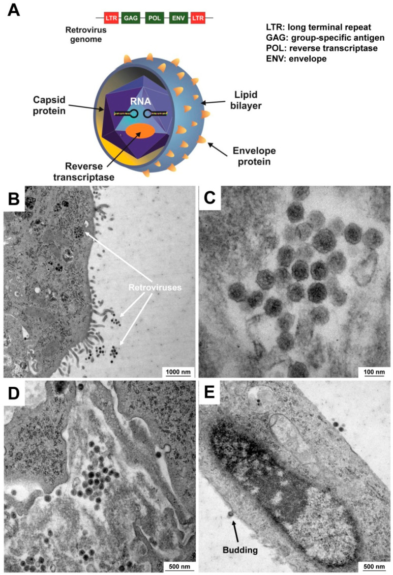
The cultivation of cells in a favorable artificial environment has become a versatile tool in cellular and molecular biology. Cultured primary cells and continuous cell lines are indispensable in investigations of basic, biomedical, and translation research. However, despite their important role, cell lines are frequently misidentified or contaminated by other cells, bacteria, fungi, yeast, viruses, or chemicals. In addition, handling and manipulating of cells is associated with specific biological and chemical hazards requiring special safeguards such as biosafety cabinets, enclosed containers, and other specialized protective equipment to minimize the risk of exposure to hazardous materials and to guarantee aseptic work conditions. This review provides a brief introduction about the most common problems encountered in cell culture laboratories and some guidelines on preventing or tackling respective problems.

Keywords: STR profiling; cell authentication; conditional reprogramming; contamination; misidentification; mycoplasma; retrovirus.


Conflict of interest statement

The authors declare no conflicts of interest.

Figures

Figure 1
Figure 1
Monitoring pH changes in cell culture by phenol red. Phenol red (Phenolsulfonphthalein) is a triphenylmethane dye used in cell cultures to monitor pH changes. Its color exhibits a gradual transition from yellow to red and fuchsia with increasing pH.
Figure 2
Figure 2
Detection of Mycoplasma spp. in cell culture supernatants. (A) Mycoplasmas are the smallest self-replicating bacteria devoid of a cell wall. They consist of a lipoprotein plasma membrane, ribosomes, and a genome consisting of a circular, double-stranded DNA molecule. (B) A monolayer of a mycoplasma-contaminated rat cell line was stained with DAPI and analyzed under a fluorescent microscope with an appropriate UV filter package (340/380 nm excitation) at a magnification of 400×. Please note the very small bright extranuclear dots that are typical for mycoplasmas (marked by arrows). The scale bar represents 50 µm. (C) In the depicted analysis, 2 µL normal control medium (Medium (−) and 2 µL medium taken from a Mycoplasma-infected cell line (Medium (+)) were tested for Mycoplasma infection using the Venor®GeM OneStep kit (#11-8050, Minerva biolabs GmbH, Berlin, Germany) according to the manufacturer’s instructions. As a positive control, 2 µL of the positive control included in the kit system was co-amplified. The internal control in each sample resulting in an amplicon of 191 bp in size demonstrates the functionality of the PCR reaction, while an amplicon of 265–278 bp in size indicates a contamination with Mycoplasma spp. The PCR products were separated on a 2% standard agarose gel containing ethidium bromide and amplicons visualized using a standard gel imager.
Figure 3
Figure 3
Selected compounds useful to remove or prevent Mycoplasma contaminations. (A) Baytril (CAS-no.: 93106-60-6), (B) Ciprobay (CAS-no.: 85721-33-1), (C) Zagam (CAS-no.: 110871-86-8), and (D) BM-Cyclin containing Minocycline hydrochloride (CAS-no.: 13614-98-7) and Tiamulin fumarate (CAS-no.: 55297-96-6) are powerful anti-mycoplasma agents that are used to eliminate or prevent mycoplasma contamination. For the biological activity of the depicted compounds, refer to Table 3.
Figure 4
Figure 4
Clearance of mycoplasma contamination in a rat cell culture as assessed by electron microcopy. A contaminated cell culture was treated with Plasmocin for 2 weeks. Shown are representative electron microscopic images of cells before (AC) and after (D) treatment with the broad-spectrum anti-mycoplasma reagent showing that the mycoplasmas were successfully eliminated from the culture. Images were taken at original magnifications of 4646× to 27,800×. Please note the characteristic fusions of the mycoplasma membrane with the cytoplasmic membrane of the host cell in (A).
Figure 5
Figure 5
Retroviral contamination in the mouse cell line GRX. (A) General structure of a retrovirus. A retrovirus is an enveloped particle about 100 nm in diameter. The envelope encoded by the env gene is composed of glycoprotein that is encapsulated by lipids obtained from the plasma membrane of the host cells during the budding process. The retroviral genome further consists of two identical single-stranded RNA molecules 7–9 kb in length. Group-specific antigens (gag) are major components of the viral capsid, while the reverse transcriptase (pol) is necessary for synthesis of viral DNA. (B) Occurrence of a not-specified retrovirus in the mouse hepatic stellate line GRX as assessed by electron microscopic analysis. Retroviral particles can be found in the culture medium and in the cytosol. (C,D) Higher magnification images of retroviruses show the typical spherical structure. (E) This electron microscopic image shows a budding process in which a retroviral particle acquires its envelope by spreading through the host’s plasma membrane. Magnifications are 10,000× (B), 100,000× (C), 27,800× (D), and 21,560× (E), respectively.
Figure 6
Figure 6
Chromatograms of typical short tandem repeat (STR) markers for a murine cell line. Genomic DNA from murine AML12 cells was isolated and subjected to STR profiling using the CellCheckTM Mouse system (IDEXX, Kornwestheim, Germany). This system amplifies 19 species-specific STR markers spread on 17 chromosomes. In this figure, four representative STR profiles from chromosomes 17, 18, 19, and chromosome X are depicted. For the complete STR profile of that cell line, refer to Table 5.

References

    1. Hubrecht R.C., Carter E. The 3Rs and humane experimental technique: Implementing change. Animals. 2019;9:754. doi: 10.3390/ani9100754. - DOI - PMC - PubMed
    1. Pamies D., Leist M., Coecke S., Bowe G., Allen D.G., Gstraunthaler G., Bal-Price A., Pistollato F., de Vries R.B.M., Hogberg H.T., et al. Guidance document on Good Cell and Tissue Culture Practice 2.0 (GCCP 2.0) ALTEX. 2022;39:30–70. doi: 10.14573/altex.2111011. - DOI - PubMed
    1. Horbach S.P.J.M., Halffman W. The ghosts of HeLa: How cell line misidentification contaminates the scientific literature. PLoS ONE. 2017;12:e0186281. doi: 10.1371/journal.pone.0186281. - DOI - PMC - PubMed
    1. Babic Z., Capes-Davis A., Martone M.E., Bairoch A., Ozyurt I.B., Gillespie T.H., Bandrowski A.E. Incidences of problematic cell lines are lower in papers that use RRIDs to identify cell lines. Elife. 2019;8:e41676. doi: 10.7554/eLife.41676. - DOI - PMC - PubMed
    1. International Cell Line Authentication Committee (ICLAC) [(accessed on 18 February 2023)]. Available online: https://iclac.org/

Publication types

LinkOut - more resources