Review
. 2023 Feb 23;12(5):708.
doi: 10.3390/cells12050708.

Role of RUNX3 in Restriction Point Regulation

Affiliations
Review

Role of RUNX3 in Restriction Point Regulation

Jung-Won Lee et al. Cells. .

Abstract

A cell cycle is a series of events that takes place in a cell as it grows and divides. At the G1 phase of cell cycle, cells monitor their cumulative exposure to specific signals and make the critical decision to pass through the restriction (R)-point. The R-point decision-making machinery is fundamental to normal differentiation, apoptosis, and G1-S transition. Deregulation of this machinery is markedly associated with tumorigenesis. Therefore, identification of the molecular mechanisms that govern the R-point decision is one of the fundamental issues in tumor biology. RUNX3 is one of the genes frequently inactivated in tumors by epigenetic alterations. In particular, RUNX3 is downregulated in most K-RAS-activated human and mouse lung adenocarcinomas (ADCs). Targeted inactivation of Runx3 in the mouse lung induces adenomas (ADs), and markedly shortens the latency of ADC formation induced by oncogenic K-Ras. RUNX3 participates in the transient formation of R-point-associated activator (RPA-RX3-AC) complexes, which measure the duration of RAS signals and thereby protect cells against oncogenic RAS. This review focuses on the molecular mechanism by which the R-point participates in oncogenic surveillance.

Keywords: K-RAS; R-point; RUNX3; cell cycle.


Conflict of interest statement

The authors declare no conflict of interest.

Figures

Figure 1
Figure 1
Fluctuation of levels of cyclins D and E and p21 through the cell cycle. The levels of cyclins D and E and p21 fluctuate markedly as cells progress through the cell cycle. The CDK inhibitors of the p21 family stimulate the formation of the cyclin D–CDK4/6 complex while inhibiting the formation of other cyclin–CDK complexes, including cyclin E–CDK2. Extracellular signals strongly influence the levels of D-type cyclins during the early G1 phase. However, the levels of the other cyclins, including cyclin E, are controlled by intracellular signals and precisely coordinated with cell cycle progression. The cyclin E–CDK2 complex is activated after cells pass through the R-point, followed by the formation of the remaining cyclin–CDK complexes through a cell-autonomous program.
Figure 2
Figure 2
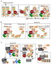
Sequential molecular events for R-point decisions. (A) In response to mitogenic stimulation, RUNX3 opens target loci. Upon mitogenic stimulation, RUNX3 binds to the enhancer regions of target loci within inactive chromatin, as indicated by H3K27-me3. pRB-E2F1 and p300 associate with RUNX3. p300 acetylates RUNX3 and histones. BRD2 binds to acetylated RUNX3 through its first bromodomain (BD1) and to H4K12-ac through its second bromodomain (BD2). Subsequently, SWI/SNF and MLL1/5 bind to the C-terminal region of BRD2. At this time point, inhibitory histones (H3K27-me3) are eliminated, and activating histones (H3K4-me3) are enriched at these loci. (B) RUNX3 forms an R-point-associated RUNX3-containing Activator (Rpa-RX3-AC) complex at the R-point. While RUNX3 binds to the enhancer region and recruits its coactivator (p300), histone-modifying enzymes (MLLs), and chromatin-remodeling complex (SWI/SNF), the basal transcription machinery (TFIID) is recruited to the promoter region of the target loci. The TFIID binds to the C-terminal region of BRD2 to form Rpa-RX3-AC. Moreover, the enhancer interacts with the promoter through Rpa-RX3-AC during the R-point. (C) Rpa-RX3-AC complex is converted to Rpa-RX3-RE after the R-point. Two hours after mitogenic stimulation, CDK4 (associated with p21) binds to RUNX3 and becomes an additional component of Rpa-RX3-AC. At this point, the cyclin D1–PRC1 complex forms separately from the Rpa-RX3-AC complex. Downregulation of the RAS-MEK signal results in the maturation of the cyclin D1–PRC1 complex in the cyclin D1–HDAC4–PRC1 complex, which binds to Rpa-RX3-AC through the interaction between cyclin D1 and CDK4, a component of the Rpa-RX3-AC complex, yielding Rpa-RX3-TR. Activation of CDK4 through its association with cyclin D1 is critical for the inactivation of the chromatin loci and the dissociation of the entire complex. RNF2, a component of the PRC2, contributes to the enrichment of an inactive chromatin marker (H2A-K119-Ub, H2A ubiquitination at Lys-119) at this locus. If the RAS signal is constitutively activated, the cyclin D1–PRC1 complex fails to mature into the cyclin D1–HDAC4-PRC1 complex, and consequently cannot form Rpa-RX3-TR. Therefore, if R-point commitment is normal, cells expressing constitutively active RAS cannot progress through the R-point into the S phase. If the mitogenic signal is downregulated in a normal manner, Rpa-RX3-TR dissociates (4 h after stimulation) into two complexes, the RUNX3–Cyclin D1–HDAC4 and BRD2–PRC1–SWI/SNF–TFIID complexes, which remain associated with chromatin. This is followed by the association of PRC2 with RUNX3–cyclin D1–HDAC4 to form Rpa-RX3-RE, which remains on the chromatin. EZH2, a component of PRC2, contributes to the enrichment of an inactive chromatin marker (H3K27-me3) at this locus.
Figure 3
Figure 3
Sequential molecular events occurring during multistep tumor progression. Most colorectal and lung adenocarcinomas develop through a multistep tumorigenesis pathway. Tumors show development from normal tissue, to adenoma (AD), to adenocarcinoma (ADC), and ultimately progress to multiple types of invasive tumors. Molecular events occurring at each step are indicated.
Figure 4
Figure 4
Inactivation of p53 tumor suppressor pathways. (A) Two major pathways trigger p53 activation. Aberrant oncogene activation is sensed by the R-point-associated complex, which induces the expression of ARF, inactivating HDM2 and stabilizing p53. DNA damage stress is sensed by the ATM/ATR kinases, activating the CHK1/CHK2 kinases, which stabilize p53. (B) Inactivation of p53 tumor suppressor pathways during multistep tumor progression. AD development is characterized by disruption of the Arf-p53 pathway due to the abrogation of the R-point, most frequently by RU3 inactivation. This may result in the selection of K-Ras-activated cells, which acquire a proliferative advantage. At the AD stage, the ATM/ATR → CHK1/2 → p53 pathway is functional. The pathway is disrupted at a late stage of AD by p53 mutation.
Figure 5
Figure 5
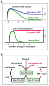
Mechanism for sensing constitutive RAS activation. (A) Normal RAS activity is downregulated to the basal level soon after mitogenic stimulation. While RAS is activated, ARF is expressed. In normal cells, ARF is expressed for only a short time (1–3 h after mitogenic stimulation), followed by its suppression when RAS activity is downregulated. However, heterozygous mutations of RAS result in the maintenance of 50% of the maximum level of RAS activity. This persistent RAS activity maintains ARF expression until the G1/S checkpoint is reached. (B) Schematic illustration of the R-point-associated oncogene surveillance mechanism. Formation of the Rpa-RX3-AC complex is triggered by the RAS-MEK pathway 1 h after serum stimulation. The complex binds to the ARF promoter through RUNX-binding sites and induces ARF expression. After the R-point (4 h after mitogenic stimulation), the RAS-MEK pathway activity is downregulated. Rpa-RX3-AC complexes are converted to Rpa-RX3-RE complexes, which suppress ARF expression. However, constitutively activated RAS signaling inhibits the conversion of Rpa-RX3-AC to Rpa-RX3-RE complexes and prolongs ARF expression, which drives cells toward apoptosis. These series of molecular events enable cells to distinguish normal mitogenic signals from abnormal oncogenic K-RAS signals, thereby constituting an R-point-associated oncogene surveillance mechanism.

References

    1. Pardee A.B. A restriction point for control of normal animal cell proliferation. Proc. Natl. Acad. Sci. USA. 1974;71:1286–1290. doi: 10.1073/pnas.71.4.1286. - DOI - PMC - PubMed
    1. Blagosklonny M.V., Pardee A.B. The restriction point of the cell cycle. Cell Cycle. 2002;1:103–110. doi: 10.4161/cc.1.2.108. - DOI - PubMed
    1. Weinberg R.A. The Biology of Cancer. Garland Science; New York, NY, USA: 2007. pp. 275–329. Chapter 8. pRB and Control of the Cell Cycle Clock.
    1. Won K.A., Xiong Y., Beach D., Gilman M.Z. Growth-regulated expression of D-type cyclin genes in human diploid fibroblasts. Proc. Natl. Acad. Sci. USA. 1992;89:9910–9914. doi: 10.1073/pnas.89.20.9910. - DOI - PMC - PubMed
    1. Sherr C.J., Roberts J.M. CDK inhibitors: Positive and negative regulators of G1-phase progression. Genes Dev. 1999;13:1501–1512. doi: 10.1101/gad.13.12.1501. - DOI - PubMed

Publication types

Substances