. 2023 Feb 23;12(5):713.
doi: 10.3390/cells12050713.

Analysis of Wild Type and Variant B Cystatin C Interactome in Retinal Pigment Epithelium Cells Reveals Variant B Interacting Mitochondrial Proteins

Affiliations

Analysis of Wild Type and Variant B Cystatin C Interactome in Retinal Pigment Epithelium Cells Reveals Variant B Interacting Mitochondrial Proteins

Emil Carlsson et al. Cells. .

Abstract

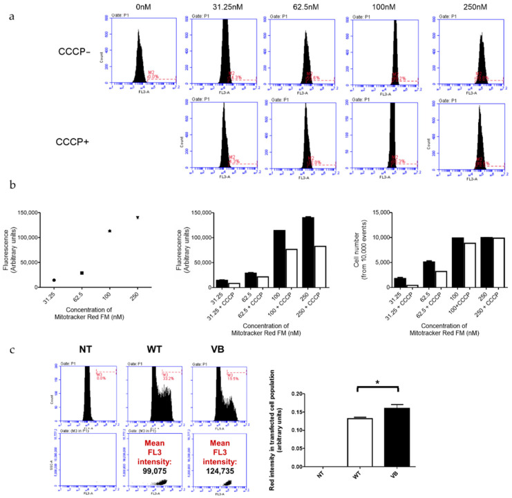
Cystatin C, a secreted cysteine protease inhibitor, is abundantly expressed in retinal pigment epithelium (RPE) cells. A mutation in the protein's leader sequence, corresponding to formation of an alternate variant B protein, has been linked with an increased risk for both age-related macular degeneration (AMD) and Alzheimer's disease (AD). Variant B cystatin C displays intracellular mistrafficking with partial mitochondrial association. We hypothesized that variant B cystatin C interacts with mitochondrial proteins and impacts mitochondrial function. We sought to determine how the interactome of the disease-related variant B cystatin C differs from that of the wild-type (WT) form. For this purpose, we expressed cystatin C Halo-tag fusion constructs in RPE cells to pull down proteins interacting with either the WT or variant B form, followed by identification and quantification by mass spectrometry. We identified a total of 28 interacting proteins, of which 8 were exclusively pulled down by variant B cystatin C. These included 18 kDa translocator protein (TSPO) and cytochrome B5 type B, both of which are localized to the mitochondrial outer membrane. Variant B cystatin C expression also affected RPE mitochondrial function with increased membrane potential and susceptibility to damage-induced ROS production. The findings help us to understand how variant B cystatin C differs functionally from the WT form and provide leads to RPE processes adversely affected by the variant B genotype.

Keywords: Alzheimer’s disease; age-related macular degeneration; aging; cystatin C; halo-tag; mistrafficking; mitochondria; translocator protein; variant B.


Conflict of interest statement

The authors declare no conflict of interest.

Figures

Figure 1
Figure 1
Expression and pull down analysis of Halo-tagged cystatin C in ARPE-19 cells. (a) WT and variant B (VB) cystatin C Halo-tag fusion constructs were expressed in ARPE-19 cells, followed by staining with Halo TMR Direct ligand and visualizing on a Zeiss Apotome fluorescence microscope. (b) Whole cell lysates of ARPE-19 cells expressing Halo-tagged WT and variant B cystatin C were analyzed by immunoblotting. Endogenously expressed cystatin C was present in all samples, alongside higher molecular weight Halo-tagged cystatin C present in the respective transfected cells. NT, non-transfected. (c) Schematic overview of workflow for pull down analysis of Halo-tagged fusion proteins.
Figure 2
Figure 2
Protein interaction analysis of WT and variant B cystatin C by mass spectrometry. (a) Silver stain analysis of eluates from Halo-tag protein pull down experiments indicate multiple proteins eluted from pull down assays using both fusion constructs. (b) Visual representation of interacting proteins identified from Halo-tag pull down analysis using WT or variant B cystatin C fusion constructs. Volcano plots of mass spectrometry results of eluates from Halo-tag pull down analysis using WT (c) or variant B (d) cystatin C fusion constructs. Proteins exclusively pulled down by variant B cystatin C marked in plot. Cells transfected with plasmid encoding Halo-tag only were used as negative controls. All experiments were conducted in biological triplicates.
Figure 3
Figure 3
Validation of protein interaction analysis of WT and variant B cystatin C by immunoblotting. Immunoblot analysis of pull down samples from ARPE-19 cells transfected with empty vector (EV), wild-type cystatin C (WT), or variant B cystatin C (VB). Top panels show inputs, middle panels show flow through (FT), and bottom panels show eluates (Elu). GAPDH was used as a negative control. IRAP (encoded by LNPEP) was pulled down by both WT and variant B cystatin C. Blots shown are representative of three separate experiments.
Figure 4
Figure 4
Analysis of mitochondrial association of variant B cystatin C and effects on mitochondrial reactive oxygen species (ROS) generation. (a) Immunoblot analysis of mitochondrial and cytoplasmic fractions of ARPE-19 cells expressing WT or variant B cystatin C Halo-tag fusion proteins. Blots were immunostained with antibodies against mitochondrial (TOM20) and cytoplasmic (α-tubulin) markers in addition to cystatin C. Blots shown are representative of three separate experiments. Graphs show normalized protein expression of Halo-tag fusion proteins in cytosolic and enriched mitochondrial fractions. (b) WT and variant B cystatin C EGFP fusion constructs were expressed in ARPE-19, followed by mitochondrial staining using Mitotracker Red FM dye and live cell analysis by fluorescence microscopy using a Zeiss Apotome microscope. Scale bars, 20 µm. (c) Mitochondrial ROS detection flow cytometry analysis in ARPE-19 cells transfected with WT and variant B cystatin EGFP fusion constructs (+/− antimycin A). After P1 gating was performed to exclude dead cells and debris (not shown), M1 (green channel) histograms were used to measure transfection efficiency (left of image). The M1 population was then selected to measure red intensity (FL2) as shown by plots (middle and right). Graphs show red fluorescence intensity (FL2 channel) in the transfected (green population; FL1 channel) cell population for basal mitochondrial ROS production (+/− antimycin A) and fold change between antimycin A treated cells/non-treated cells for each condition (arbitrary units ± S.E.M. minimum of n = 3; Student’s t test * p ≤ 0.05, *** p ≤ 0.001).
Figure 5
Figure 5
Analysis of mitochondrial membrane potential (Δψm) in ARPE-19 cells transfected with WT and variant B (VB) cystatin C EGFP fusion constructs. (a) Evaluation of optimal Mitotracker Red FM concentration required to stain the entire ARPE-19 cell population (+/− CCCP, an uncoupling agent that disrupts oxidative phosphorylation). Concentrations used for optimization ranged between 0–250 nM. Flow cytometry utilizing the FL3 channel was used for analysis. (b) Graphs show FL3 fluorescence intensity for cells stained with different concentrations of Mitotracker Red FM (left = optimization line graph; middle = +/− CCCP optimization bar graph; and right = number of cells stained with the concentration range of Mitotracker Red FM +/− CCCP). (c) Flow cytometry analysis of ARPE-19 cells transfected with EGFP WT or variant B cystatin and stained with Mitotracker Red FM. After P1 gating was performed to exclude dead cells and debris (not shown), M1 (green channel) histograms were used to measure transfection efficiency (left). The M1 population was then selected to measure red intensity (FL3), as shown by middle and right plots. Graph shows red fluorescence intensity (FL3 channel) in the transfected (green; FL1 channel) cell population (arbitrary units ± S.E.M. minimum of n = 3; Student’s t test * p ≤ 0.05).

References

    1. Pennington K.L., DeAngelis M.M. Epidemiology of age-related macular degeneration (AMD): Associations with cardiovascular disease phenotypes and lipid factors. Eye Vision. 2016;3:34. doi: 10.1186/s40662-016-0063-5. - DOI - PMC - PubMed
    1. Ambati J., Fowler B.J. Mechanisms of age-related macular degeneration. Neuron. 2012;75:26–39. doi: 10.1016/j.neuron.2012.06.018. - DOI - PMC - PubMed
    1. Kinnunen K., Petrovski G., Moe M.C., Berta A., Kaarniranta K. Molecular mechanisms of retinal pigment epithelium damage and development of age-related macular degeneration. Acta Ophthalmol. 2012;90:299–309. doi: 10.1111/j.1755-3768.2011.02179.x. - DOI - PubMed
    1. Fritsche L.G., Fariss R.N., Stambolian D., Abecasis G.R., Curcio C.A., Swaroop A. Age-related macular degeneration: Genetics and biology coming together. Annu. Rev. Genomics Hum. Genet. 2014;15:151–171. doi: 10.1146/annurev-genom-090413-025610. - DOI - PMC - PubMed
    1. Fritsche L.G., Igl W., Bailey J.N., Grassmann F., Sengupta S., Bragg-Gresham J.L., Burdon K.P., Hebbring S.J., Wen C., Gorski M., et al. A large genome-wide association study of age-related macular degeneration highlights contributions of rare and common variants. Nat. Genet. 2016;48:134–143. doi: 10.1038/ng.3448. - DOI - PMC - PubMed

Publication types

MeSH terms

LinkOut - more resources