

Human Bone Marrow-Derived Mesenchymal Stromal Cells Reduce the Severity of Experimental Necrotizing Enterocolitis in a Concentration-Dependent Manner
- PMID: 36899900
- PMCID: PMC10000931
- DOI: 10.3390/cells12050760
Human Bone Marrow-Derived Mesenchymal Stromal Cells Reduce the Severity of Experimental Necrotizing Enterocolitis in a Concentration-Dependent Manner
Abstract
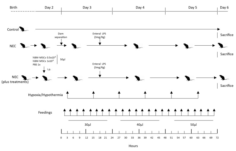
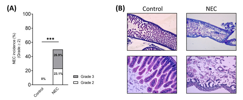
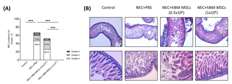
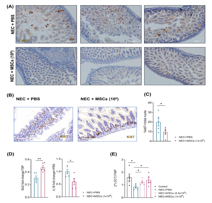
Necrotizing enterocolitis (NEC) is a devastating gut disease in preterm neonates. In NEC animal models, mesenchymal stromal cells (MSCs) administration has reduced the incidence and severity of NEC. We developed and characterized a novel mouse model of NEC to evaluate the effect of human bone marrow-derived MSCs (hBM-MSCs) in tissue regeneration and epithelial gut repair. NEC was induced in C57BL/6 mouse pups at postnatal days (PND) 3-6 by (A) gavage feeding term infant formula, (B) hypoxia/hypothermia, and (C) lipopolysaccharide. Intraperitoneal injections of PBS or two hBM-MSCs doses (0.5 × 10⁶ or 1 × 10⁶) were given on PND2. At PND 6, we harvested intestine samples from all groups. The NEC group showed an incidence of NEC of 50% compared with controls (p < 0.001). Severity of bowel damage was reduced by hBM-MSCs compared to the PBS-treated NEC group in a concentration-dependent manner, with hBM-MSCs (1 × 10⁶) inducing a NEC incidence reduction of up to 0% (p < 0.001). We showed that hBM-MSCs enhanced intestinal cell survival, preserving intestinal barrier integrity and decreasing mucosal inflammation and apoptosis. In conclusion, we established a novel NEC animal model and demonstrated that hBM-MSCs administration reduced the NEC incidence and severity in a concentration-dependent manner, enhancing intestinal barrier integrity.
Keywords: apoptosis; caspase 3; human bone marrow mesenchymal stromal cells; inflammation; interleukin 1b; mouse model; necrotizing enterocolitis; neonate; zonula occludens-1.
Conflict of interest statement
The authors declare that the research was conducted without any commercial or financial relationships that could be construed as a potential conflict of interest.
Figures
Similar articles
-
Cell-based therapies in preclinical models of necrotizing enterocolitis: a systematic review and meta-analysis.Stem Cells Transl Med. 2025 Feb 11;14(2):szae102. doi: 10.1093/stcltm/szae102. Stem Cells Transl Med. 2025. PMID: 40036304 Free PMC article.
-
Mesenchymal stem cell therapy in necrotizing enterocolitis: a rat study.Pediatr Res. 2011 Nov;70(5):489-94. doi: 10.1203/PDR.0b013e31822d7ef2. Pediatr Res. 2011. PMID: 21772224
-
Administration of extracellular vesicles derived from human amniotic fluid stem cells: a new treatment for necrotizing enterocolitis.Pediatr Surg Int. 2021 Mar;37(3):301-309. doi: 10.1007/s00383-020-04826-6. Epub 2021 Feb 10. Pediatr Surg Int. 2021. PMID: 33566163
-
Treatment of experimental necrotizing enterocolitis with stem cell-derived exosomes.J Pediatr Surg. 2018 Jun;53(6):1215-1220. doi: 10.1016/j.jpedsurg.2018.02.086. Epub 2018 Mar 14. J Pediatr Surg. 2018. PMID: 29661576 Free PMC article.
-
The Impact of MicroRNAs in Neonatal Necrotizing Enterocolitis and other Inflammatory Conditions of Intestine: A Review.Curr Pediatr Rev. 2022;19(1):5-14. doi: 10.2174/1573396318666220117102119. Curr Pediatr Rev. 2022. PMID: 35040406 Review.
Cited by
-
Transcriptomic analysis of BM-MSCs identified EGR1 as a transcription factor to fully exploit their therapeutic potential.Biochim Biophys Acta Mol Cell Res. 2024 Dec;1871(8):119818. doi: 10.1016/j.bbamcr.2024.119818. Epub 2024 Aug 19. Biochim Biophys Acta Mol Cell Res. 2024. PMID: 39168411 Free PMC article.
-
Cell-based therapies in preclinical models of necrotizing enterocolitis: a systematic review and meta-analysis.Stem Cells Transl Med. 2025 Feb 11;14(2):szae102. doi: 10.1093/stcltm/szae102. Stem Cells Transl Med. 2025. PMID: 40036304 Free PMC article.
-
Oral administration of bone marrow-derived mesenchymal stem cells attenuates intestinal injury in necrotizing enterocolitis.Clin Exp Pediatr. 2024 Mar;67(3):152-160. doi: 10.3345/cep.2023.01151. Epub 2024 Feb 19. Clin Exp Pediatr. 2024. PMID: 38369803 Free PMC article.
-
Advances in neonatal cell therapies: Proceedings of the First Neonatal Cell Therapies Symposium (2022).Pediatr Res. 2023 Nov;94(5):1631-1638. doi: 10.1038/s41390-023-02707-x. Epub 2023 Jun 28. Pediatr Res. 2023. PMID: 37380752 Free PMC article. Review.
-
Bone marrow-derived mesenchymal stromal cells in necrotizing enterocolitis treatment: a narrative review.Front Pediatr. 2025 Jul 31;13:1624236. doi: 10.3389/fped.2025.1624236. eCollection 2025. Front Pediatr. 2025. PMID: 40822681 Free PMC article. Review.
