Review
. 2023 Mar 4;12(5):804.
doi: 10.3390/cells12050804.

Polyunsaturated Fatty Acids Drive Lipid Peroxidation during Ferroptosis

Affiliations
Review

Polyunsaturated Fatty Acids Drive Lipid Peroxidation during Ferroptosis

Michael S Mortensen et al. Cells. .

Abstract

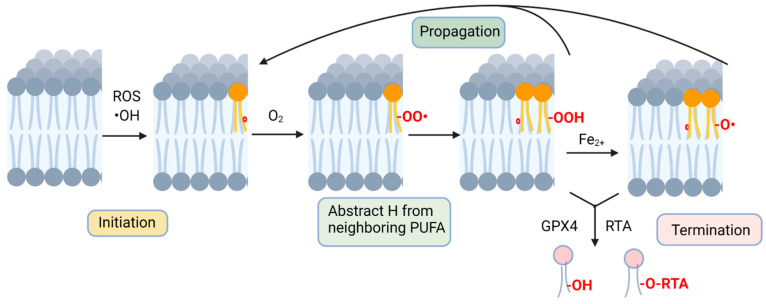
Ferroptosis is a form of regulated cell death that is intricately linked to cellular metabolism. In the forefront of research on ferroptosis, the peroxidation of polyunsaturated fatty acids has emerged as a key driver of oxidative damage to cellular membranes leading to cell death. Here, we review the involvement of polyunsaturated fatty acids (PUFAs), monounsaturated fatty acids (MUFAs), lipid remodeling enzymes and lipid peroxidation in ferroptosis, highlighting studies revealing how using the multicellular model organism Caenorhabditis elegans contributes to the understanding of the roles of specific lipids and lipid mediators in ferroptosis.

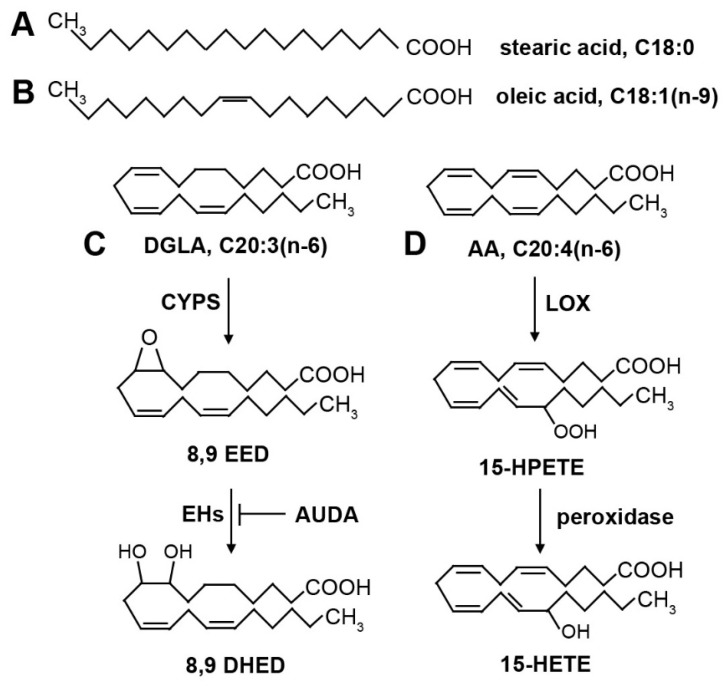
Keywords: AA; Caenorhabditis elegans; DGLA; MUFA; PUFA; ferroptosis; lipid peroxidation.


Conflict of interest statement

The authors declare no conflict of interest.

Figures

Figure 1
Figure 1
Non-enzymatic lipid peroxidation. Peroxidation is initiated by cellular ROS, where hydroxyl, alkoxyl or peroxyl radicals abstract a hydrogen from a PUFA acyl group (radical electrons denoted as red circle). A PUFA peroxide is formed by reacting with molecular oxygen and abstraction of a hydrogen from an adjacent membrane PUFA. Fenton chemistry contributes to further lipid radical formation, contributing to the chain reaction of lipid radicals attacking acyl groups on nearby unsaturated phospholipid molecules. Lipid peroxidation is terminated by actions of radical-trapping antioxidants or by reduction by catalyzed by glutathione peroxidase activity. Figure created with BioRender.com, accessed on 1 February 2023.
Figure 2
Figure 2
Structures of common fatty acids and oxygenated derivatives. (A) Stearic acid (18:0) is a saturated fatty acid. (B) Oleic acid (18:1n-9) is a monounsaturated fatty acid. The single double bond is in the cis position, creating a kink in the fatty acid that prevents tight packing of fatty acids and contributes to membrane fluidity. (C) Dihommo-γ linolenic acid (DGLA, 20:3n-6) is a polyunsaturated fatty acid. Oxygenated derivatives are produced by cytochrome P450 (CYP) enzymes, forming an epoxide. The double bond that is converted to an epoxide depends on the position-specific isoform of CYP enzymes. The epoxides can be converted into diols by epoxide hydrolase (EH) enzymes. The EH enzymes are inhibited by AUDA. (D) Arachidonic acid (AA, 20:4n-6) is a polyunsaturated fatty acid. Shown are examples of oxygenated derivatives produced by lipoxygenase (LOX) enzymes and peroxidase activity. The location of the hydroperoxide is dependent on the position-specific isoform of LOX. The hydroperoxide can be further reduced by peroxidase activity, leading to a bioactive hydroxyl derivative.
Figure 3
Figure 3
Dietary DGLA causes ferroptosis of germ cells and sterility in C. elegans. (A) Schematic of the C. elegans fatty acid supplementation assay. Synchronized L1 larvae are plated onto agar plates containing DGLA and dietary E. coli, and incubated at 20 degrees until they reach adulthood, when they are scored as fertile or sterile. Sterile worms lack gametes due to ferroptosis of germ cells during development. (B) Mutant strains that are more sensitive to DGLA are known as enhancers, while mutant strains that are less sensitive to DGLA are known as suppressors. Often, enhancer strains contain mutations in protective genes, such as genes encoding GPX enzymes or genes required for MUFA production. Suppressor genes include genes needed to produce membrane PUFAs, or mutants that confer increased stress responses.

References

    1. Dixon S.J., Lemberg K.M., Lamprecht M.R., Skouta R., Zaitsev E.M., Gleason C.E., Patel D.N., Bauer A.J., Cantley A.M., Yang W.S., et al. Ferroptosis: An Iron-Dependent Form of Nonapoptotic Cell Death. Cell. 2012;149:1060–1072. doi: 10.1016/j.cell.2012.03.042. - DOI - PMC - PubMed
    1. Stockwell B.R. Ferroptosis turns 10: Emerging mechanisms, physiological functions, and therapeutic applications. Cell. 2022;185:2401–2421. doi: 10.1016/j.cell.2022.06.003. - DOI - PMC - PubMed
    1. Lee J.-Y., Kim W., Bae K.-H., Lee S., Lee E.-W. Lipid Metabolism and Ferroptosis. Biology. 2021;10:184. doi: 10.3390/biology10030184. - DOI - PMC - PubMed
    1. Dixon S.J., Stockwell B.R. The Hallmarks of Ferroptosis. Annu. Rev. Cancer Biol. 2019;3:35–54. doi: 10.1146/annurev-cancerbio-030518-055844. - DOI
    1. Jiang X., Stockwell B.R., Conrad M. Ferroptosis: Mechanisms, biology and role in disease. Nat. Rev. Mol. Cell Biol. 2021;22:266–282. doi: 10.1038/s41580-020-00324-8. - DOI - PMC - PubMed

Publication types

Substances

LinkOut - more resources