Review
. 2023 Feb 28;15(5):1509.
doi: 10.3390/cancers15051509.

Technical Implications for Surgical Resection in Locally Advanced Pancreatic Cancer

Affiliations
Review

Technical Implications for Surgical Resection in Locally Advanced Pancreatic Cancer

Martín de Santibañes et al. Cancers (Basel). .

Abstract

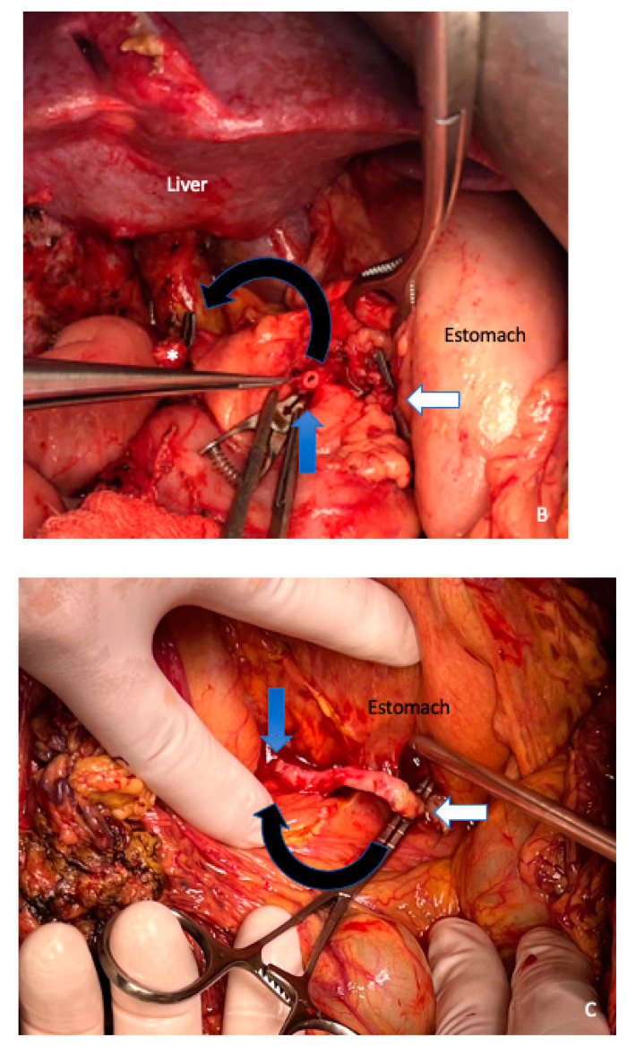
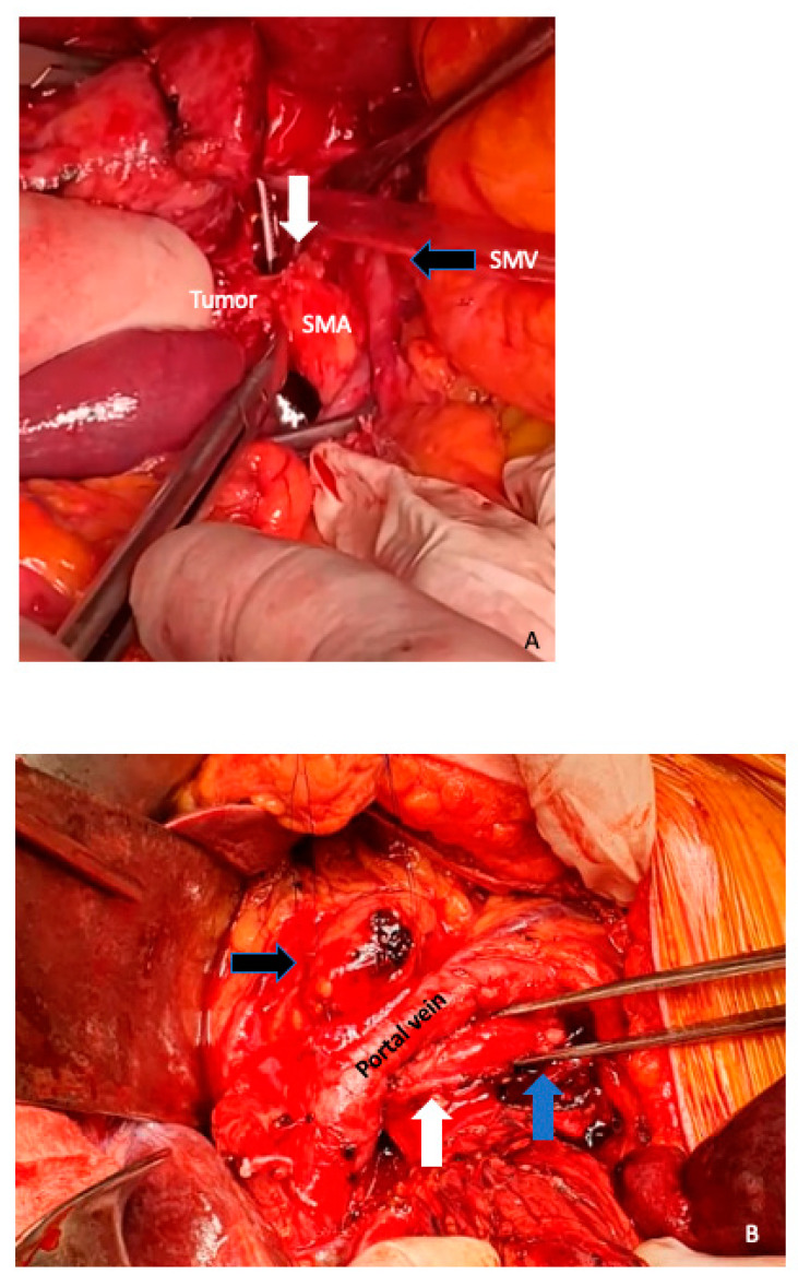
Pancreatic ductal adenocarcinoma remains a global health challenge and is predicted to soon become the second leading cause of cancer death in developed countries. Currently, surgical resection in combination with systemic chemotherapy offers the only chance of cure or long-term survival. However, only 20% of cases are diagnosed with anatomically resectable disease. Neoadjuvant treatment followed by highly complex surgical procedures has been studied over the last decade with promising short- and long-term results in patients with locally advanced pancreatic ductal adenocarcinoma (LAPC). In recent years, a wide variety of complex surgical techniques that involve extended pancreatectomies, including portomesenteric venous resection, arterial resection, or multi-organ resection, have emerged to optimize local control of the disease and improve postoperative outcomes. Although there are multiple surgical techniques described in the literature to improve outcomes in LAPC, the comprehensive view of these strategies remains underdeveloped. We aim to describe the preoperative surgical planning as well different surgical resections strategies in LAPC after neoadjuvant treatment in an integrated way for selected patients with no other potentially curative option other than surgery.

Keywords: arterial resection; extended pancreatectomies; locally advanced pancreatic ductal adenocarcinoma; neoadjuvant treatment; portomesenteric venous resection.


Conflict of interest statement

None of the authors of this manuscript have any direct or indirect commercial financial incentive associated with the publication of this paper.

Figures

Figure 1
Figure 1
A 67 year old female patient with jaundice and abdominal pain. Endoscopic ultrasound biopsy confirmed poorly differentiated PDAC and a biliary stent was placed (asterisk). MDCT shows expansive pancreatic formation located in the cephalic portion, with poorly defined limits, measuring approximately 54 × 32 mm (A). Exophytic tumor (T), encompassing the SMA and distal branches (white arrow) and the spleno–mesenteric–portal confluence, extending towards the superior mesenteric vein (encasement) (B,C). Cephalically it extends towards the celiac trunk, encompassing its terminal branches (black arrow) (D).
Figure 1
Figure 1
A 67 year old female patient with jaundice and abdominal pain. Endoscopic ultrasound biopsy confirmed poorly differentiated PDAC and a biliary stent was placed (asterisk). MDCT shows expansive pancreatic formation located in the cephalic portion, with poorly defined limits, measuring approximately 54 × 32 mm (A). Exophytic tumor (T), encompassing the SMA and distal branches (white arrow) and the spleno–mesenteric–portal confluence, extending towards the superior mesenteric vein (encasement) (B,C). Cephalically it extends towards the celiac trunk, encompassing its terminal branches (black arrow) (D).
Figure 2
Figure 2
In extensive Kocher maneuver in combination with the Cattell–Braasch mobilizations of the cecum, right colon, right colonic flexure, and the root of the small bowel, together with the Treitz ligament, an adequate exposure of the entire infrahepatic vena cava (IVC), aorta, and the left renal vein (white arrow) is achieved.
Figure 3
Figure 3
Identification of the superior mesenteric vessels (the blue arrow shows the superior mesenteric artery) is performed at the root of the mesentery below the transverse mesocolon to the origin from the aorta. The middle colic artery is detached at its origin from the SMA in this manner (white arrow), and the tumor-infiltrated section of the transverse mesocolon is resected to remain with the specimen.
Figure 4
Figure 4
(A) Radical antegrade modular pancreatosplenectomy (RAMPS) proceeds in a right-to-left antegrade manner, with early parenchymal transection at the neck of the pancreas, close to the gastroduodenal artery (blue arrow) and early control of the splenic vessels (in its origin) (white arrow). The black arrow marks the common hepatic artery, and the asterisk shows a partial venous excision spleno–mesenteric confluence with direct closure (venorrhaphy). (B) Posterior RAMPS. The posterior magnitude of dissection was behind the left adrenal gland, including the Gerota fascia and the fat tissue around the left kidney. The left renal artery is shown with white arrow. Blue arrow shows the pancreas stump. Black arrow, superior mesenteric vein.
Figure 5
Figure 5
The TRIANGLE operation (blue triangle zone) is a proper approach to achieve a complete and radical removal of the tumor and associated lymphatic or perineural extension along a region defined anatomically by the origins of the celiac trunk (white arrow), the superior mesenteric artery (SMA) (blue arrow), the portal vein (anteriorly), and superior mesenteric vein (SMV).
Figure 6
Figure 6
(A) Autologous parietal peritoneum was harvested from the right hypochondrium. (B) Type 2 vein reconstruction, using falciform ligament patch over the spleno–portal junction (white circle). The asterisk shows the distal pancreatic stump. The blue arrow shows the superior mesenteric artery. PV, portal vein. SMV, superior mesenteric vein.
Figure 6
Figure 6
(A) Autologous parietal peritoneum was harvested from the right hypochondrium. (B) Type 2 vein reconstruction, using falciform ligament patch over the spleno–portal junction (white circle). The asterisk shows the distal pancreatic stump. The blue arrow shows the superior mesenteric artery. PV, portal vein. SMV, superior mesenteric vein.
Figure 7
Figure 7
Type 3: segmental superior mesenteric vein resection (white arrow) with primary veno-venous anastomosis. The blue arrow shows the splenic vein. The asterisk, the distal pancreatic stump. SMA, superior mesenteric artery.
Figure 8
Figure 8
Type 4: segmental resection with interposed venous conduit. The spleno–portal confluent was complete resected. The blue arrow shows the superior mesenteric vein. The blue lines mark the cadaveric iliac vein interposed conduit of 6.5 cm long. SMA, superior mesenteric artery, which was clamped for 25 min to perform the venous anastomosis and avoid intestinal congestion.
Figure 9
Figure 9
Venous jump graft technique. (A) Identification of the superior mesenteric vein (white asterisk) at the root of the mesentery (blue arrow). (B) Anastomosis between the superior mesenteric vein (blue arrow) and cadaveric iliac venous bank graft (black asterisk). The white arrow marks the distal segment that will be anastomosed with the portal vein or a collateral vessel. The jump graft is accessed through the transverse mesocolon (black arrow).
Figure 10
Figure 10
Splenic artery transposition technique with respective anastomosis with proper hepatic artery. (A) The right angle shows the splenic artery being dissected from its origin in the celiac trunk (blue arrow) distally (white arrow). The black arrow shows proper hepatic artery (bulldog clamp). (B) Section towards the distal portion of the splenic artery. The white arrow shows the distal end of the splenic artery that will be ligated. The blue arrow, the portion of the splenic artery that will be anastomosed to the proper hepatic artery (asterisk). The black arrow is marking the 180° rotation that the splenic artery will undergo to achieve a tension-free anastomosis. (C) Anastomosis between the splenic artery and proper hepatic artery. The white arrow is marking the clamp in the proximal segment of the splenic artery. The black arrow shows the 4 cm segment of the splenic artery that was rotated 180°, with the respective anastomosis to the proper hepatic artery (blue arrow).
Figure 10
Figure 10
Splenic artery transposition technique with respective anastomosis with proper hepatic artery. (A) The right angle shows the splenic artery being dissected from its origin in the celiac trunk (blue arrow) distally (white arrow). The black arrow shows proper hepatic artery (bulldog clamp). (B) Section towards the distal portion of the splenic artery. The white arrow shows the distal end of the splenic artery that will be ligated. The blue arrow, the portion of the splenic artery that will be anastomosed to the proper hepatic artery (asterisk). The black arrow is marking the 180° rotation that the splenic artery will undergo to achieve a tension-free anastomosis. (C) Anastomosis between the splenic artery and proper hepatic artery. The white arrow is marking the clamp in the proximal segment of the splenic artery. The black arrow shows the 4 cm segment of the splenic artery that was rotated 180°, with the respective anastomosis to the proper hepatic artery (blue arrow).
Figure 11
Figure 11
(A) Arterial divestment, is achieved by placing it in a plane between the uninvolved arterial wall and the tumor tissue of the affected arterial segment. The dissection plane should be placed between the periarterial nerve plexus and the arterial adventitia. We usually perform this maneuver with cold scissors to avoid thermal damage to the arterial wall, as can be seen in the image. The white arrow shows the limit between the tumor and the superior mesenteric artery (SMA) wall. Black arrow, retracted superior mesenteric vein (SMV) (B) The white arrow is showing the tunica adventitia of the superior mesenteric artery, which has then been removed distally, to give a tumor margin: the tunica media of the superior mesenteric artery can be seen at the level of the forceps and marked with a blue arrow. Pancreas stump (black arrow).

References

    1. Siegel R.L., Miller K.D., Jemal A. Cancer statistics. CA Cancer J. Clin. 2018;68:7–30. doi: 10.3322/caac.21442. - DOI - PubMed
    1. Klaiber U., Schnaidt E.S., Hinz U., Gaida M.M., Heger U., Hank T., Strobel O., Neoptolemos J.P., Mihaljevic A.L., Büchler M.W., et al. Prognostic Factors of Survival After Neoadjuvant Treatment and Resection for Initially Unresectable Pancreatic Cancer. Ann. Surg. 2021;273:154–162. doi: 10.1097/SLA.0000000000003270. - DOI - PubMed
    1. Amin M.B., Greene F.L., Edge S.B., Compton C.C., Gershenwald J.E., Brookland R.K., Meyer L., Gress D.M., Byrd D.R., Winchester D.P. The Eighth Edition AJCC Cancer Staging Manual: Continuing to build a bridge from a population-based to a more “personalized” approach to cancer staging. CA Cancer J. Clin. 2017;67:93–99. doi: 10.3322/caac.21388. - DOI - PubMed
    1. Bockhorn M., Uzunoglu F.G., Adham M., Imrie C., Milicevic M., Sandberg A.A., Asbun H.J., Bassi C., Büchler M., Charnley R.M., et al. International Study Group of Pancreatic Surgery. Borderline resectable pancreatic cancer: A consensus statement by the International Study Group of Pancreatic Surgery (ISGPS) Surgery. 2014;155:977–988. doi: 10.1016/j.surg.2014.02.001. - DOI - PubMed
    1. Kirkegård J., Aahlin E.K., Al-Saiddi M., Bratlie S.O., Coolsen M., de Haas R.J., den Dulk M., Fristrup C., Harrison E.M., Mortensen M.B., et al. Multicentre study of multidisciplinary team assessment of pancreatic cancer resectability and treatment allocation. Br. J. Surg. 2019;106:756–764. doi: 10.1002/bjs.11093. - DOI - PubMed

LinkOut - more resources