. 2023 Mar 3;20(5):4523.
doi: 10.3390/ijerph20054523.

Ellagic Acid Prevents Particulate Matter-Induced Pulmonary Inflammation and Hyperactivity in Mice: A Pilot Study

Affiliations

Ellagic Acid Prevents Particulate Matter-Induced Pulmonary Inflammation and Hyperactivity in Mice: A Pilot Study

Sunyoung Jeong et al. Int J Environ Res Public Health. .

Abstract

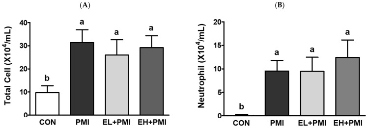
The inhalation of fine particulate matter (PM) is a significant health-related environmental issue. Previously, we demonstrated that repeated PM exposure causes hyperlocomotive activity in mice, as well as inflammatory and hypoxic responses in their lungs. In this study, we evaluated the potential efficacy of ellagic acid (EA), a natural polyphenolic compound, against PM-induced pulmonary and behavioral abnormalities in mice. Four treatment groups were assigned in this study (n = 8): control (CON), particulate-matter-instilled (PMI), low-dose EA with PMI (EL + PMI), and high-dose EA with PMI (EH + PMI). EA (20 and 100 mg/kg body weight for low dose and high dose, respectively) was orally administered for 14 days in C57BL/6 mice, and after the eighth day, PM (5 mg/kg) was intratracheally instilled for 7 consecutive days. PM exposure induced inflammatory cell infiltration in the lungs following EA pretreatment. Moreover, PM exposure induced inflammatory protein expression in the bronchoalveolar lavage fluid and the expression of inflammatory (tumor necrosis factor alpha (Tnfα), interleukin (Il)-1b, and Il-6) and hypoxic (vascular endothelial growth factor alpha (Vegfα), ankyrin repeat domain 37 (Ankrd37)) response genes. However, EA pretreatment markedly prevented the induction of expression of inflammatory and hypoxic response genes in the lungs. Furthermore, PM exposure significantly triggered hyperactivity by increasing the total moving distance with an increase in moving speed in the open field test. On the contrary, EA pretreatment significantly prevented PM-induced hyperactivity. In conclusion, dietary intervention with EA may be a potential strategy to prevent PM-induced pathology and activity.

Keywords: ellagic acid; hyperactivity; hypoxia; inflammation; particulate matter.


Conflict of interest statement

The authors declare no conflict of interest.

Figures

Figure 1
Figure 1
Body and relative weight changes in mice after ellagic acid (EA) pretreatment and particulate matter (PM) instillation. C57BL/6 mice were orally pretreated with ellagic acid (EL; 20 mg/kg or EH; 100 mg/kg) for 7 days, and then PM (5 mg/kg) was intratracheally administrated for 7 days with or without ellagic acid pretreatment: (A) body weight, (B) delta BW (final minus initial BW), (C) liver/body weight ratio, and (D) lung/body weight ratio. Values represent the mean ± standard deviation (SD) for each group (n = 8). Based on the D’Agostino and Pearson omnibus test, one-way ANOVA with Tukey’s post hoc test (for (AC)) or Kruskal–Wallis test with Dunn’s post hoc test (for (D)) was applied. Lowercase letters indicate significant differences. p < 0.05.
Figure 1
Figure 1
Body and relative weight changes in mice after ellagic acid (EA) pretreatment and particulate matter (PM) instillation. C57BL/6 mice were orally pretreated with ellagic acid (EL; 20 mg/kg or EH; 100 mg/kg) for 7 days, and then PM (5 mg/kg) was intratracheally administrated for 7 days with or without ellagic acid pretreatment: (A) body weight, (B) delta BW (final minus initial BW), (C) liver/body weight ratio, and (D) lung/body weight ratio. Values represent the mean ± standard deviation (SD) for each group (n = 8). Based on the D’Agostino and Pearson omnibus test, one-way ANOVA with Tukey’s post hoc test (for (AC)) or Kruskal–Wallis test with Dunn’s post hoc test (for (D)) was applied. Lowercase letters indicate significant differences. p < 0.05.
Figure 2
Figure 2
Histological changes in lung tissue after ellagic acid (EA) pretreatment and particulate matter (PM) instillation. C57BL/6 mice were orally pretreated with ellagic acid (EL; 20 mg/kg or EH; 100 mg/kg) for 7 days, and then PM (5 mg/kg) was intratracheally administrated for 7 days with or without ellagic acid pretreatment. Representative images of hematoxylin and eosin (H & E) stained sections obtained from the (A) CON, (B) PMI, (C) EL + PMI, and (D) EH + PMI groups (n = 8). Blue arrows indicate black-pigment-laden macrophages in the alveoli and alveolar lumen interstitium. Red arrows point to the infiltrated inflammatory cells in the peribronchiolar, perivascular, and interstitial regions. Scale bars = 100 μm.
Figure 3
Figure 3
Inflammatory cell profiles in the bronchoalveolar lavage fluid (BALF) after ellagic acid (EA) pretreatment and particulate matter (PM) instillation. C57BL/6 mice were orally pretreated with ellagic acid (EL; 20 mg/kg or EH; 100 mg/kg) for 7 days, and then PM (5 mg/kg) was intratracheally administrated for 7 days with or without ellagic acid pretreatment. All the cells in the BALF were stained with Diff-Quik solution and counted: (A) total cells, (B) neutrophils, (C) macrophages, (D) eosinophils, and (E) lymphocytes. Values represent the mean ± standard deviation (SD) for each group (n = 8). Based on the D’Agostino and Pearson omnibus test, one-way ANOVA with Tukey’s post hoc test (for (AC,E)) or Kruskal–Wallis test with Dunn’s post hoc test (for (D)) was applied. Lowercase letters indicate significant differences. p < 0.05.
Figure 3
Figure 3
Inflammatory cell profiles in the bronchoalveolar lavage fluid (BALF) after ellagic acid (EA) pretreatment and particulate matter (PM) instillation. C57BL/6 mice were orally pretreated with ellagic acid (EL; 20 mg/kg or EH; 100 mg/kg) for 7 days, and then PM (5 mg/kg) was intratracheally administrated for 7 days with or without ellagic acid pretreatment. All the cells in the BALF were stained with Diff-Quik solution and counted: (A) total cells, (B) neutrophils, (C) macrophages, (D) eosinophils, and (E) lymphocytes. Values represent the mean ± standard deviation (SD) for each group (n = 8). Based on the D’Agostino and Pearson omnibus test, one-way ANOVA with Tukey’s post hoc test (for (AC,E)) or Kruskal–Wallis test with Dunn’s post hoc test (for (D)) was applied. Lowercase letters indicate significant differences. p < 0.05.
Figure 4
Figure 4
Inflammatory cytokine and H2O2 levels in the BALF after ellagic acid (EA) pretreatment and particulate matter (PM) instillation. C57BL/6 mice were orally pretreated with ellagic acid (EL; 20 mg/kg or EH; 100 mg/kg) for 7 days, and then PM (5 mg/kg) was intratracheally administrated for 7 days with or without ellagic acid pretreatment: (A) TNFα, (B) IL-6, and (C) H2O2 levels in the BALF. Values represent the mean ± standard deviation (SD) for each group (n = 8). Data passed the D’Agostino and Pearson omnibus normality test; one-way ANOVA with Tukey’s post hoc test was applied. Lowercase letters indicate significant differences. p < 0.05.
Figure 5
Figure 5
Pulmonary mRNA expressions after ellagic acid (EA) pretreatment and particulate matter (PM) instillation. C57BL/6 mice were orally pretreated with ellagic acid (EL; 20 mg/kg or EH; 100 mg/kg) for 7 days, and then PM (5 mg/kg) was intratracheally administrated for 7 days with or without ellagic acid pretreatment: (A) Tnfα, (B) Il-1β, (C) Il-6, (D) Vegfα, and (E) Ankrd37 expression was analyzed using qRT-PCR normalized to Gapdh mRNA expression. Values represent the mean ± standard deviation (SD) for each group (n = 8). Data passed the D’Agostino and Pearson omnibus normality test; one-way ANOVA with Tukey’s post hoc test was applied. Lowercase letters indicate significant differences. p < 0.05.
Figure 6
Figure 6
Open field test results and serum corticosterone levels after ellagic acid (EA) pretreatment and particulate matter (PM) instillation. C57BL/6 mice were orally pretreated with ellagic acid (EL; 20 mg/kg or EH; 100 mg/kg) for 7 days, and then PM (5 mg/kg) was intratracheally administrated for 7 days with or without ellagic acid pretreatment: (A) total distance moved, (B) distance moved in border, (C) distance moved in center, (D) number of entries into center, (E) time spent in border, (F) time spent in center, (G) the total mean speed, (H) mean speed in border, (I) mean speed in center, (J) max speed in border, (K) max speed in center of mice within 10 min of the open field test, and (L) serum corticosterone measured 1 day after the OFT. Values represent the mean ± standard deviation (SD) for each group (n = 8). Data passed the D’Agostino and Pearson omnibus test; one-way ANOVA with Tukey’s post hoc test was applied. Lowercase letters indicate significant differences. p < 0.05.
Figure 6
Figure 6
Open field test results and serum corticosterone levels after ellagic acid (EA) pretreatment and particulate matter (PM) instillation. C57BL/6 mice were orally pretreated with ellagic acid (EL; 20 mg/kg or EH; 100 mg/kg) for 7 days, and then PM (5 mg/kg) was intratracheally administrated for 7 days with or without ellagic acid pretreatment: (A) total distance moved, (B) distance moved in border, (C) distance moved in center, (D) number of entries into center, (E) time spent in border, (F) time spent in center, (G) the total mean speed, (H) mean speed in border, (I) mean speed in center, (J) max speed in border, (K) max speed in center of mice within 10 min of the open field test, and (L) serum corticosterone measured 1 day after the OFT. Values represent the mean ± standard deviation (SD) for each group (n = 8). Data passed the D’Agostino and Pearson omnibus test; one-way ANOVA with Tukey’s post hoc test was applied. Lowercase letters indicate significant differences. p < 0.05.

References

    1. Wichmann H.E. Diesel exhaust particles. Inhal. Toxicol. 2007;19((Suppl. 1)):241–244. doi: 10.1080/08958370701498075. - DOI - PubMed
    1. Ichinose T., Furuyama A., Sagai M. Biological effects of diesel exhaust particles (DEP). II. Acute toxicity of DEP introduced into lung by intratracheal instillation. Toxicology. 1995;99:153–167. doi: 10.1016/0300-483X(94)03013-R. - DOI - PubMed
    1. Iwai K., Adachi S., Takahashi M., Moller L., Udagawa T., Mizuno S., Sugawara I. Early oxidative DNA damages and late development of lung cancer in diesel exhaust-exposed rats. Environ. Res. 2000;84:255–264. doi: 10.1006/enrs.2000.4072. - DOI - PubMed
    1. Nemmar A., Al-Salam S., Zia S., Yasin J., Al Husseni I., Ali B.H. Diesel exhaust particles in the lung aggravate experimental acute renal failure. Toxicol. Sci. 2010;113:267–277. doi: 10.1093/toxsci/kfp222. - DOI - PubMed
    1. Morsi A.A., Fouad H., Alasmari W.A., Faruk E.M. The biomechanistic aspects of renal cortical injury induced by diesel exhaust particles in rats and the renoprotective contribution of quercetin pretreatment: Histological and biochemical study. Environ. Toxicol. 2022;37:310–321. doi: 10.1002/tox.23399. - DOI - PubMed