. 2023 Feb 21;24(5):4273.
doi: 10.3390/ijms24054273.

Clinical-Scale Mesenchymal Stem Cell-Derived Extracellular Vesicle Therapy for Wound Healing

Affiliations

Clinical-Scale Mesenchymal Stem Cell-Derived Extracellular Vesicle Therapy for Wound Healing

Jieun Kim et al. Int J Mol Sci. .

Abstract

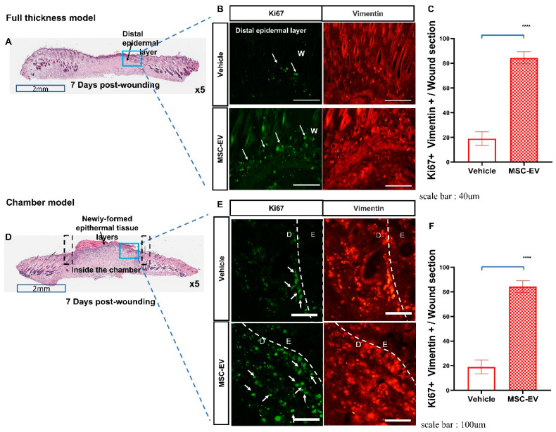
We developed an extracellular vesicle (EV) bioprocessing platform for the scalable production of human Wharton's jelly mesenchymal stem cell (MSC)-derived EVs. The effects of clinical-scale MSC-EV products on wound healing were tested in two different wound models: subcutaneous injection of EVs in a conventional full-thickness rat model and topical application of EVs using a sterile re-absorbable gelatin sponge in the chamber mouse model that was developed to prevent the contraction of wound areas. In vivo efficacy tests showed that treatment with MSC-EVs improved the recovery following wound injury, regardless of the type of wound model or mode of treatment. In vitro mechanistic studies using multiple cell lines involved in wound healing showed that EV therapy contributed to all stages of wound healing, such as anti-inflammation and proliferation/migration of keratinocytes, fibroblasts, and endothelial cells, to enhance wound re-epithelialization, extracellular matrix remodeling, and angiogenesis.

Keywords: exosomes; extracellular vesicles; functional recovery; mesenchymal stem cells; wound healing.


Conflict of interest statement

The authors declare no conflict of interest.

Figures

Figure 1
Figure 1
Characterization of extracellular vesicles (EVs) obtained from 3D culture system. (A) EVs imaged using electron microscopy (TEM and Cryo-EM); (B) histogram representing the size distributions and concentrations of the EVs using NanoSight. (C) The tetraspanins, including CD9, CD63, CD81, using Exoview analysis; and (D,E) EV positive markers, including CD63, CD81, syntenin-1 (enzyme-linked immunosorbent assay, ELISA) and EV negative markers, including histone H2A.Z and GM130 (Western blot). (F) Change in EVs according to storage period at RT.
Figure 2
Figure 2
Evaluation of cutaneous wound healing in a conventional full-thickness rat wound model (upper panel) and a chamber mouse wound model (lower panel). (A) Schematic diagram of a full-thickness wound healing model (B) Representative image of a wound over 14 d. (C) Rate of wound closure (%, re-epithelialization) measured via histo-morphometric analysis of tissue sections. (D,E) Photomicrographs of hematoxylin and eosin (H&E)-stained histologic sections of wounded skin (Scanscope image, USA) 14 d post-wounding. Graph of measurements in panel (E). (F) Schematic diagram of the time schedule of the wound chamber model. (G) Surgical processes for the mouse skin excisional wound model. (H) Workflow for evaluating the mouse skin chamber wound model. (I) Increase in both wound closure percentage and the ratio of the area of granulation tissue to the wound field was observed in EV-treated mice. (J) Photo data of wound healing 1 d after chamber removal. (K) Semiquantitative evaluation of the gap width. Measurement of wound area percentage in the chamber. Thickness of the newly formed epidermis and measurement of the wound area inside the chamber. The number of animals used at each time point was six. Data represent the mean ± standard deviation (SD). **** p < 0.0001, ** p < 0.01.
Figure 3
Figure 3
EVs promote the proliferation and migration of keratinocytes during the initial epidermal hypertrophy process. (A,B) Mesenchymal stem cell (MSC)-EVs accelerate wound healing with individually migrating keratinocytes in a mouse chamber model. Scale bar: 40 μm. (C) H&E-stained histopathological whole-slide images of full-thickness wounds at 3, 7, 10, 14, and 21 d post-wounding. (Unwounded, day 3, magnification, 100×, scale bar, 100 μm), (days post-wounding, from day 7 to 14, magnification, 4×, scale bar, 2 mm), (days post-wounding, from day 21, magnification, 100×, scale bar, 100 μm). (D) Serial changes in epidermal thickness were measured and compared between the EV and control groups. (E) Immunohistochemical image of the epidermis and (F) measurement of epidermal thickness 7 d after wound induction. White arrows indicate Ki67+/keratinocyte cells. Scale bar: 100 m. Data represent the mean ± SD. ns: p > 0.05, not significant, **** p < 0.0001, ** p < 0.01, and * p < 0.05.
Figure 4
Figure 4
EVs promote fibroblast cell proliferation and migration. (A,D) Representative cross-sectional images of H&E-stained mouse dorsal skin with epidermal structures of MSC-EV-treated wounds. Areas marked with black lines represent the Chambers, blue boxes indicate the newly created granulation tissue, and downward arrows indicate the newly formed epidermis. Scale bar: 2 mm. (B,E) Ki-67-positive fibroblast cells (GFP-TRITC merged cells) in typical cross-sections of distal epidermal layer of the vehicle group or MSC-EV-treated group. (Panels (A,D): magnification, 5×, scale bar: 2 mm), (Panel (B): Full thickness wound model, scale bar: 40 μm), (Panel (E): Chamber model, scale bar: 100 μm). D: Dermal tissue; E: Epidermal tissue; W: wound. (C,F) Quantification of the percentage of fibroblasts expressing the Ki-67 antigen/wound areas. Data represent the mean ± SD. **** p < 0.0001.
Figure 5
Figure 5
Effects of EVs on angiogenesis in the chamber wound model. (A) Immunohistochemical staining for CD31 (a marker of vascular structure) in the dermis and neovascularization sites at the center of the wound. Arrow heads indicate CD31 + vessels. Left panel, scale bar: 100 μm; Right panel, scale bar: 40 μm (B) Quantification of CD31+ cells and vessels per 20× field in the wound bed at the center of wounds treated with the vehicle or MSC-EVs collected on day 7 post-wounding. (C) On day 14, vascular endothelial growth factor (VEGF)-positive blood vessels were observed in MSC-EV-injected wounds. Arrow heads indicate VEGF + vessels. Scale bar: 40 μm (D) Quantification of VEGF+ cells and vessels per 20× field in the wound bed at the center of wounds treated with the vehicle or MSC-EVs collected on day 14 post-wounding. (EG) ELISA analyses of angiogenesis protein expression in MSC-EV-treated wound and vehicle groups. Each sample was assessed in duplicate, and the analysis was conducted thrice independently. Error bars indicate the mean ± SD. p-values were calculated using an unpaired Student’s t-test. **** p < 0.0001, *** p < 0.001, ** p < 0.01, and * p < 0.05; ns: p > 0.05, not significant.
Figure 6
Figure 6
In vitro assay for MSC-EV effects on four major cell types involved in wound healing: keratinocytes, fibroblasts, endothelial cells, and inflammatory cells. (A,B) Scratch wound healing, tube formation, and release of VEGF in vitro assays. Scratched HaCaT (A) and NIH-3T3 (B) cell monolayer treated with MSC-EVs and imaged after 24 h. Wound closure area was determined via ImageJ software analysis. Data are presented as the means of three independent measurements. Magnification: ×10. *** p < 0.001, ** p < 0.01, and ns: p > 0.05, not significant. compared to the vehicle group. Scale bar 400 μm. (C) Human umbilical vein endothelial cells (HUVECs) cultured in a Matrigel-coated 96-well plate were incubated with different doses of EVs for 24 h. Representative images of HUVEC tube formation are shown, and endothelial tube formation in each group was quantified. Scale bar 500 μm. (D) RAW264.7 cells were pretreated with various concentrations (2, 5, and 10 × 108/mL) of EVs with lipopolysaccharide (LPS; 200 ng/mL) for 24 h. Cell morphology was visualized via optical microscopy (Scale bar 100 μm, Panel (a)). Data are expressed as the mean ± standard error of the mean (SEM) of three independent experiments (Panel (b)). Percentage viability of RAW264.7 cells untreated (normal group), treated with only 200 ng/mL LPS (positive control), and treated with various particle numbers of MSC-EVs in the presence of 200 ng/mL LPS for 24 h. Data are represented as the mean ± SD of three independent experiments. Scale bar 100 μm (E) Macrophage polarization. To examine the effects of MSC-EVs on macrophage differentiation, we treated RAW264.7 cells in the presence or absence of LPS with traditional M1 and M2 macrophage-differentiated cytokines followed by flow cytometry analysis with CD80 and CD206 surface markers. MSC-EVs dominantly promoted macrophage cells toward CD206+ (M2 marker), not CD80+ (M1 marker) cell differentiation on RAW264.7 cells (E). Scale bar 40 μm. Panel (a): Microscopic images depicting the morphology of cells at different stages of the polarization protocol. Panel (b): M2 macrophages stimulated with increasing concentrations of EVs in 200 ng/mL LPS. (F) Quantitative analysis of pro-inflammatory (IL-6, IL-1b, and tumor necrosis factor-α) and anti-inflammatory cytokines (IL-10) in mouse skin wounds via ELISA. Data are represented as the mean ± SD. The results are representative of three experiments. Student’s t-test was used for analysis. **** p < 0.0001, *** p < 0.001, ** p < 0.01, and ns: p > 0.05, not significant.

References

    1. Doi H., Kitajima Y., Luo L., Yan C., Tateishi S., Ono Y., Urata Y., Goto S., Mori R., Masuzaki H., et al. Potency of umbilical cord blood- and Wharton’s jelly-derived mesenchymal stem cells for scarless wound healing. Sci. Rep. 2016;6:18844. doi: 10.1038/srep18844. - DOI - PMC - PubMed
    1. Maxson S., Lopez E.A., Yoo D., Danilkovitch-Miagkova A., Leroux M.A. Concise review: Role of mesenchymal stem cells in wound repair. Stem Cells Transl. Med. 2012;1:142–149. doi: 10.5966/sctm.2011-0018. - DOI - PMC - PubMed
    1. Badiavas E.V., Falanga V. Treatment of chronic wounds with bone marrow-derived cells. Arch Derm. 2003;139:510–516. doi: 10.1001/archderm.139.4.510. - DOI - PubMed
    1. Vojtassak J., Danisovic L., Kubes M., Bakos D., Jarabek L., Ulicna M., Blasko M. Autologous biograft and mesenchymal stem cells in treatment of the diabetic foot. Neuro Endocrinol. Lett. 2006;27((Suppl. S2)):134–137. - PubMed
    1. Squillaro T., Peluso G., Galderisi U. Clinical Trials With Mesenchymal Stem Cells: An Update. Cell Transpl. 2016;25:829–848. doi: 10.3727/096368915X689622. - DOI - PubMed

LinkOut - more resources