Review
. 2023 Feb 22;24(5):4360.
doi: 10.3390/ijms24054360.

Recent Advances and Progress on Melanin: From Source to Application

Affiliations
Review

Recent Advances and Progress on Melanin: From Source to Application

Lili Guo et al. Int J Mol Sci. .

Abstract

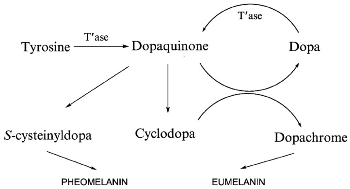
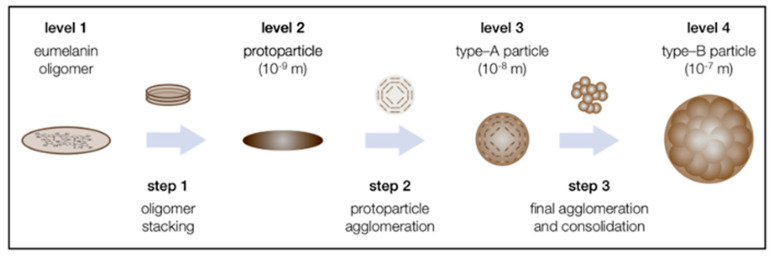
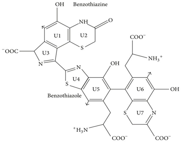
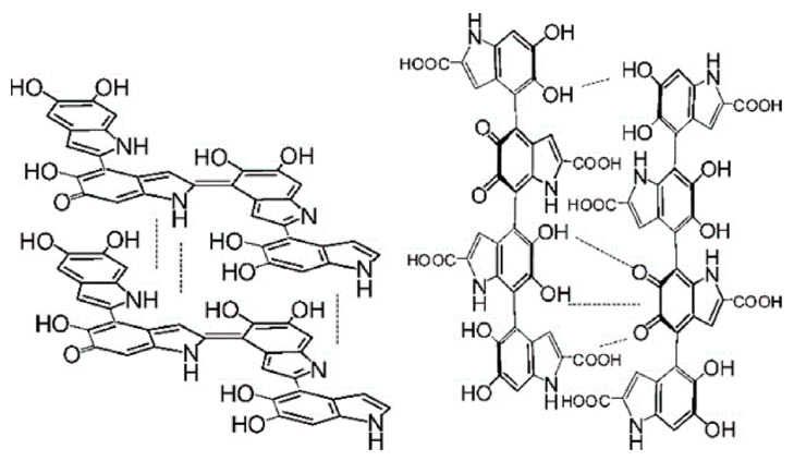
Melanin is a biological pigment formed by indoles and phenolic compounds. It is widely found in living organisms and has a variety of unique properties. Due to its diverse characteristics and good biocompatibility, melanin has become the focus in the fields of biomedicine, agriculture, the food industry, etc. However, due to the wide range of melanin sources, complex polymerization properties, and low solubility of specific solvents, the specific macromolecular structure and polymerization mechanism of melanin remain unclear, which significantly limits the further study and application of melanin. Its synthesis and degradation pathways are also controversial. In addition, new properties and applications of melanin are constantly being discovered. In this review, we focus on the recent advances in the research of melanin in all aspects. Firstly, the classification, source, and degradation of melanin are summarized. Secondly, a detailed description of the structure, characterization, and properties of melanin is followed. The novel biological activity of melanin and its application is described at the end.

Keywords: application; bioactivity; decomposition; melanin; structure; synthesis.


Conflict of interest statement

The authors declare no conflict of interest.

Figures

Figure 1
Figure 1
Divergence of melanogenic pathways. Adapted from [57] with minor modifications.
Figure 2
Figure 2
The proposed three-step mechanism for the supramolecular buildup of eumelanin based on the state of the literature and the presented results. Adapted from [56] with minor modifications.
Figure 3
Figure 3
Structural diagram of benzothiazine and benzothiazole. Adapted from [3] with minor modifications.
Figure 4
Figure 4
Representative structures of DHI and DHICA melanin. Left: DHI, right: DHICA. Adapted from [75] with minor modifications.
Figure 5
Figure 5
In vitro antibacterial activities are tested using (a) E. coli and (c) S. aureus. Statistically significant differences are indicated by *** p < 0.001, ** p < 0.01, and * p < 0.05 compared to the control. Representative images of agar plates are shown after incubating (b) E. coli and (d) S. aureus in melanin (150 mg mL−1) for 4 h at 37 °C. Reprinted with permission from Ref. [175]. Copyright 2021 Royal Society of Chemistry.
Figure 6
Figure 6
Escherichia coli (E. coli) and Staphylococcus aureus (S. aureus) were incubated with H-melanin and T-melanin, followed by NIR irradiation for 15 min. Photographs show E. coli and S. aureus after incubation with melanin, followed by NIR irradiation. Reprinted with permission from Ref. [177]. Copyright 2020 Elsevier.
Figure 7
Figure 7
The illustration depicts 1-induced autophagy sensitizing photothermal killing of cancer cells. PPBR was internalized by cancer cells via RGD-αvβ3recognition. Beclin 1 on the nanoparticle surface up-regulated autophagy to sensitize cancer cells to PTT. Reprinted with permission from Ref. [34]. Copyright 2019 Elsevier.

References

    1. Borovanský J. Melanins and Melanosomes. John Wiley & Sons; Hoboken, NJ, USA: 2011. History of Melanosome Research; pp. 1–19.
    1. Pralea I., Moldovan R., Petrache A., Ilies M., Hegheș S.-C., Ielciu I., Nicoara R., Moldovan M., Ene M., Radu M., et al. From Extraction to Advanced Analytical Methods: The Challenges of Melanin Analysis. Int. J. Mol. Sci. 2019;20:3943. doi: 10.3390/ijms20163943. - DOI - PMC - PubMed
    1. Solano F. Melanins: Skin Pigments and Much More—Types, Structural Models, Biological Functions, and Formation Routes. New J. Sci. 2014;2014:498276. doi: 10.1155/2014/498276. - DOI
    1. Caldas M., Paiva-Santos A.C., Veiga F., Rebelo R., Reis R.L., Correlo V. Melanin nanoparticles as a promising tool for biomedical applications-a review. Acta Biomater. 2020;105:26–43. doi: 10.1016/j.actbio.2020.01.044. - DOI - PubMed
    1. Morison W.L. What Is the Function of Melanin? Arch. Dermatol. 1985;121:1160–1163. doi: 10.1001/archderm.1985.01660090074017. - DOI - PubMed

LinkOut - more resources