. 2023 Feb 23;24(5):4418.
doi: 10.3390/ijms24054418.

Malonyl-CoA Accumulation as a Compensatory Cytoprotective Mechanism in Cardiac Cells in Response to 7-Ketocholesterol-Induced Growth Retardation

Affiliations

Malonyl-CoA Accumulation as a Compensatory Cytoprotective Mechanism in Cardiac Cells in Response to 7-Ketocholesterol-Induced Growth Retardation

Mei-Ling Cheng et al. Int J Mol Sci. .

Abstract

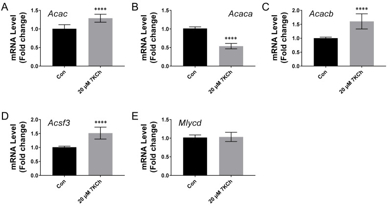
The major oxidized product of cholesterol, 7-Ketocholesterol (7KCh), causes cellular oxidative damage. In the present study, we investigated the physiological responses of cardiomyocytes to 7KCh. A 7KCh treatment inhibited the growth of cardiac cells and their mitochondrial oxygen consumption. It was accompanied by a compensatory increase in mitochondrial mass and adaptive metabolic remodeling. The application of [U-13C] glucose labeling revealed an increased production of malonyl-CoA but a decreased formation of hydroxymethylglutaryl-coenzyme A (HMG-CoA) in the 7KCh-treated cells. The flux of the tricarboxylic acid (TCA) cycle decreased, while that of anaplerotic reaction increased, suggesting a net conversion of pyruvate to malonyl-CoA. The accumulation of malonyl-CoA inhibited the carnitine palmitoyltransferase-1 (CPT-1) activity, probably accounting for the 7-KCh-induced suppression of β-oxidation. We further examined the physiological roles of malonyl-CoA accumulation. Treatment with the inhibitor of malonyl-CoA decarboxylase, which increased the intracellular malonyl-CoA level, mitigated the growth inhibitory effect of 7KCh, whereas the treatment with the inhibitor of acetyl-CoA carboxylase, which reduced malonyl-CoA content, aggravated such a growth inhibitory effect. Knockout of malonyl-CoA decarboxylase gene (Mlycd-/-) alleviated the growth inhibitory effect of 7KCh. It was accompanied by improvement of the mitochondrial functions. These findings suggest that the formation of malonyl-CoA may represent a compensatory cytoprotective mechanism to sustain the growth of 7KCh-treated cells.

Keywords: 7-ketocholesterol; malonyl-CoA; mevalonic acid.


Conflict of interest statement

The authors declare no conflict of interest. The funders had no role in the design of the study; in the collection, analyses, or interpretation of data; in the writing of the manuscript, or in the decision to publish the results.

Figures

Figure 1
Figure 1
7KCh inhibits the growth of cardiomyocytes. HL-1 (A) or AC16 (B) cells were seeded at a cell density of 5 × 10⁴ per well of a 24 well culture plate, and after attachment, they were treated with the indicated concentrations of 7KCh for 24 h. Cells were stained with Hoechst 33342. The cell number was quantified using IN Cell Analyzer 1000 and is expressed as the percentage relative to the untreated cells. Data are mean ± SD of six experiments. *** p < 0.005, **** p < 0.001 vs. the 0 μM 7KCh treatment group.
Figure 2
Figure 2
Mitochondrial dysfunction in cardiomyocytes is caused by 7KCh. HL-1 (AI) and AC16 (JR) cells were treated with the indicated concentrations of 7KCh for 24 h and stained with MitoSOX red (A,J) or JC1 (B,K). The mean fluorescence intensity (MFI) of the MitoSOX red-stained cells is expressed as the percentage relative to that of the untreated control (A,J). The ratio of the MFI of FL2 channel to that of FL1 channel (i.e., JC1 FL2/FL1 ratio) was calculated and is expressed as the percentage relative to that of the untreated control (B,K). Data are mean ± SD, N = 12. ** p < 0.01, *** p < 0.001, **** p < 0.005 vs. the 0 μM 7KCh treatment group. (C,L) The HL-1 cells or AC16 cells were treated with the indicated concentrations of 7KCh for 24 h and subjected to Seahorse respirometry analysis. Oligomycin, FCCP, rotenone, and antimycin were injected at appropriate timepoints. The oxygen consumption rate (OCR) and extracellular acidification rate (ECAR) were assessed. The OCR is normalized to cell number and plotted as a function of time of the 7KCh-treated cells (C,L). The baseline OCR and ECAR of the cells which were treated with the vehicle (Con) or with the indicated concentrations of 7KCh are normalized to cell number and plotted on the energy map (D,M). A representative of three experiments is shown. The basal respiration (E,N), maximum respiration (F,O), spare respiratory capacity (G,P), and proton leak (H,Q) were calculated. (I,R) Cells were similarly treated and extracted for ATP quantification. The ATP level is expressed as the percentage relative to that of the untreated control. Data are mean ± SD of six experiments. * p < 0.05, ** p < 0.01, *** p < 0.001, **** p < 0.005 vs. the 0 μM 7KCh treatment group.
Figure 3
Figure 3
Compensatory mitochondrial biogenesis is induced by 7KCh. (A,B) HL-1 cells were treated with the indicated concentrations of 7KCh and analyzed for the expression of porin and actin by immunoblotting with respective antibodies. A representative of three experiments is shown. The band intensities of porin and actin were quantified. The band intensity of porin was normalized to that of actin and is expressed as the fold change relative to the 0 μM 7KCh treatment group. Data are mean ± SD of three experiments. * p < 0.05 vs. the 0 μM 7KCh treatment group. (C) Cells were similarly treated, and the mitochondrial mass was analyzed by MitoTracker Green staining and flow cytometric analysis. The MFI of the MitoTracker Green-stained cells is expressed as the fold change relative to that of the untreated control. Data are mean ± SD, N = 9. * p < 0.05 vs. the 0 μM 7KCh treatment group. (D,E) Expression of characteristic respiratory complex proteins. HL-1 cells were treated with the indicated concentrations of 7KCh, and analyzed for expression of NDUFB8, SDHB, UQCRC2, ATP5F1A, MTCO1, and glyceraldehyde-3-phosphate dehydrogenase (GAPDH). A representative of three experiments is shown. The band intensities of various proteins were quantified. The band intensities of these proteins are normalized to that of GAPDH, and expressed as the fold change relative to the 0 μM 7KCh treatment group. Data are mean ± SD of three experiments. * p < 0.05 vs. the 0 μM 7KCh treatment group.
Figure 4
Figure 4
The reprogramming of cardiomyocytic metabolism and malonyl-CoA accumulation is induced by 7KCh. (A,B) The HL-1 cells were treated with the vehicle (Control) or with the indicated concentrations of 7KCh for 24 h, and subsequently, harvested for LC-MS/MS analysis in the electrospray ionization (ESI) negative ion mode. The OPLS-DA score plot (A) and the heatmap showing metabolites with significant differences between the 7KCh treatment group versus the control (B) are shown. The percent relative abundance is color-coded. Data are mean ± SD of six experiments. (C) A schematic diagram of the 7KCh treatment and [U-13C] glucose labeling procedure is shown. HL-1 (D) and AC16 (E) cells were treated with the vehicle or with 10 or 20 μM 7KCh for 24 h, labeled with [U-13C] glucose for 1 h, and harvested for MS and isotopologue analysis. Distribution of M (blue), M+2 (i.e., with 2 13C atoms; green), M+3 (orange), M+4 (grey), M+5 (pink) and M+6 (red) isotopologues of metabolites were analyzed using the liquid chromatography/time-of-flight/mass spectrometry (LC-TOF-MS) system. The abundances of the selected metabolites (malonyl-CoA, acetyl-CoA, HMG-CoA, FBP, citrate, succinyl-CoA, and OAA) are expressed as the percentage relative to the 0 μM 7KCh treatment group (Con). The histograms are mapped onto the biochemical pathways (D). (E) The levels of HMG-CoA, malonyl-CoA and acetyl-CoA in AC16 cells treated with the indicated concentrations of 7KCh are shown. Data are mean ± SD of six experiments.
Figure 5
Figure 5
CPT-1 activity and β-oxidation in cardiomyocytes are inhibited by 7KCh. (A,B) The HL-1 cells were treated with the vehicle (Con) or 20 μM 7KCh for 24 h, and a crude mitochondrial fraction was prepared for the analysis of CPT-1 and porin expression by immunoblotting (A) and for the measurement of CPT-1 activity (B). For immunoblotting (A), a representative of three experiments is shown. (B) The CPT-1 activity of the 7KCh-treated cells is expressed as the percentage of that of untreated control cells. Data are mean ± SD, N = 6. ** p < 0.01 vs. the untreated control cells. (C) The crude mitochondrial fraction of the HL-1 cells was treated with the vehicle (Con) or 5 μM malonyl-CoA and assayed for the CPT-1 activity. Data are mean ± SD, N = 6. The CPT-1 activity of the malonyl-CoA treatment group is expressed as the percentage of that of Con group. **** p < 0.005 vs. the Con group. (D) The HL-1 cells were treated with the vehicle (Con) or 20 μM 7KCh for 24 h and were subjected to fatty acid oxidation assay using palmitate-BSA as a substrate (or BSA as the vehicle control for palmitate treatment; vehicle). A representative of three experiments is shown. (E) The cells were similarly treated, and the total RNA was extracted for the qRT-PCR-based quantification of Cpt1a, Cpt1b, and Cpt1c expression. Data are mean ± SD, N = 3.
Figure 6
Figure 6
Accumulation of malonyl-CoA partially reverses the growth inhibitory effect of 7KCh. (A) A schematic diagram shows the enzymatic reactions at which pharmacological inhibitors act. CBM 301940 is an inhibitor of malonyl-CoA decarboxylase (red stop sign); ND 646 is an inhibitor of acetyl-CoA carboxylase (green stop sign); lovastatin is an inhibitor of HMGCR (blue stop sign). (BD) The HL-1 cells were seeded at a cell density of 5 × 10⁴ per well of a 24-well culture plate, and after attachment, they were treated with the vehicle (vehicle) or with 0.25, 1, or 10 μM lovastatin (B), with the vehicle (vehicle) or with 0.5, 2.5, or 10 μM CBM 301940 (C), or with the vehicle (vehicle) or with 25, 100 or 250 nM ND 646 (D), in addition to 0 μM (i.e., the DMSO vehicle control; solid bar) or 20 μM 7KCh (striped bar) for 24 h. The cells were stained with Hoechst 33342, and the cell number was quantified using IN Cell Analyzer 1000. Data are mean ± SD of six experiments. * p < 0.05, ** p < 0.01, *** p < 0.005 vs. the vehicle group for the respective inhibitor treatment. (EG) The cells were similarly treated with 7KCh and the pharmacological inhibitors and extracted for the quantification of HMG-CoA, malonyl-CoA, and acetyl-CoA. The abundances of these metabolites are expressed as the percentage relative to those of the cells that were treated with neither 7KCh nor any pharmacological inhibitor. Data are mean ± SD, N = 3. * p < 0.05, ** p < 0.01, *** p < 0.005, the 7KCh treatment group vs. the non-treatment group; # p < 0.05, ## p < 0.01, ### p < 0.005 vs. the vehicle group for the respective inhibitor treatment.
Figure 7
Figure 7
Knockout of the Mlycd gene restores mitochondrial functions and counteracts the growth inhibitory effect of 7KCh. (A) The Mlycd−/− and control AC16 cells were analyzed for the expression of MLYCD and GAPDH by immunoblotting. A representative of three experiments is shown. (B) The Mlycd−/− and control AC16 cells were treated with 0 (i.e., the vehicle control) or 20 μM 7KCh for 24 h and stained with Hoechst 33342. The cell number was quantified using IN Cell Analyzer 1000 and is expressed as the percentage relative to the untreated AC16 cells. Data are mean ± SD of six experiments. * p < 0.05 vs. AC16 cells. (C,D) The Mlycd−/− and control AC16 cells were treated with the indicated concentrations of 7KCh for 24 h and stained with JC1 (C) or MitoSOX red (D). The ratio of the MFI of FL2 channel to that of FL1 channel (i.e., JC1 FL2/FL1 ratio) was calculated and is expressed as the percentage relative to that of the untreated AC16 cells. The MFI of the MitoSOX red-stained cells is expressed as the percentage relative to that of the untreated AC16 cells. Data are mean ± SD, N = 6. ** p < 0.01 vs. AC16 cells. (EG) The Mlycd−/− and control AC16 cells were similarly treated with 7KCh and extracted for the quantification of HMG-CoA, malonyl-CoA, and acetyl-CoA. The abundances of these metabolites are expressed as the percentage relative to those of the untreated AC16 cells. Data are mean ± SD, N = 6. * p < 0.05, ** p < 0.01, *** p < 0.005 vs. AC16 cells.
Figure 8
Figure 8
The transcription of the genes encoding malonyl-CoA metabolizing enzymes is regulated by 7KCh. The HL-1 cells were treated with 0 (i.e., the vehicle control; Con) or 20 μM 7KCh, and the total RNA was extracted for the qRT-PCR-based quantification of expression of the Acac (A) (i.e., all transcript variants encoding different acetyl-CoA carboxylase isoforms), Acaca (B), Acacb (C), Acsf3 (D), and Mlycd (E) genes. The abundances of these metabolites are expressed as the fold change relative to the untreated control. Data are mean ± SD, N = 12. **** p < 0.001 vs. the untreated control.

References

    1. Brown A.J., Jessup W. Oxysterols and atherosclerosis. Atherosclerosis. 1999;142:1–28. doi: 10.1016/S0021-9150(98)00196-8. - DOI - PubMed
    1. Schweizer R.A., Zurcher M., Balazs Z., Dick B., Odermatt A. Rapid hepatic metabolism of 7-ketocholesterol by 11beta-hydroxysteroid dehydrogenase type 1: Species-specific differences between the rat, human, and hamster enzyme. J. Biol. Chem. 2004;279:18415–18424. doi: 10.1074/jbc.M313615200. - DOI - PubMed
    1. Lyons M.A., Samman S., Gatto L., Brown A.J. Rapid hepatic metabolism of 7-ketocholesterol in vivo: Implications for dietary oxysterols. J. Lipid Res. 1999;40:1846–1857. doi: 10.1016/S0022-2275(20)34901-4. - DOI - PubMed
    1. Lee J.W., Huang J.D., Rodriguez I.R. Extra-hepatic metabolism of 7-ketocholesterol occurs by esterification to fatty acids via cPLA2alpha and SOAT1 followed by selective efflux to HDL. Biochim. Biophys. Acta. 2015;1851:605–619. doi: 10.1016/j.bbalip.2015.01.007. - DOI - PMC - PubMed
    1. Tang H.Y., Wang C.H., Ho H.Y., Wu P.T., Hung C.L., Huang C.Y., Wu P.R., Yeh Y.H., Cheng M.L. Lipidomics reveals accumulation of the oxidized cholesterol in erythrocytes of heart failure patients. Redox Biol. 2018;14:499–508. doi: 10.1016/j.redox.2017.10.020. - DOI - PMC - PubMed

LinkOut - more resources