Review
. 2023 Feb 24;24(5):4523.
doi: 10.3390/ijms24054523.

COVID-19 Biogenesis and Intracellular Transport

Affiliations
Review

COVID-19 Biogenesis and Intracellular Transport

Alexander A Mironov et al. Int J Mol Sci. .

Abstract

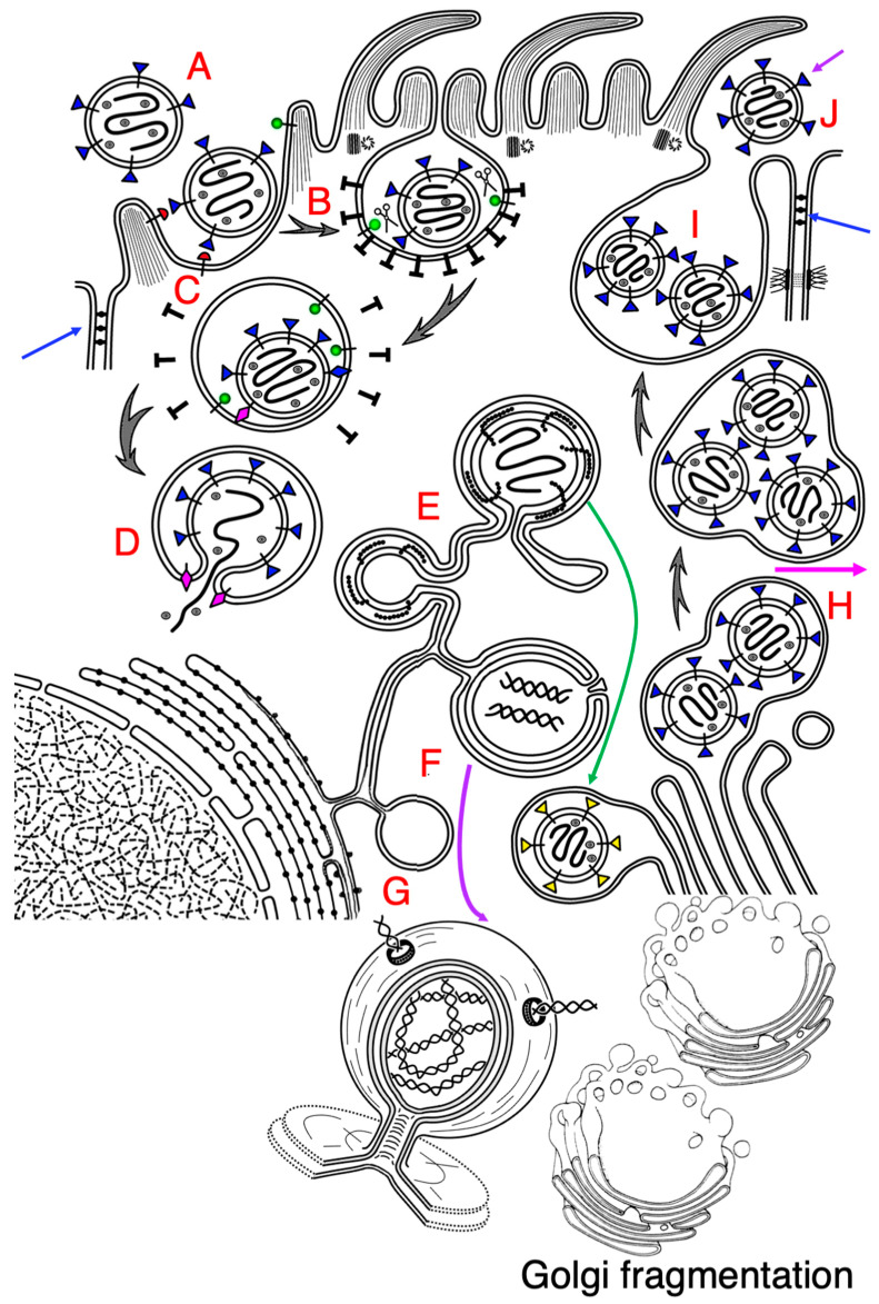
SARS-CoV-2 is responsible for the COVID-19 pandemic. The structure of SARS-CoV-2 and most of its proteins of have been deciphered. SARS-CoV-2 enters cells through the endocytic pathway and perforates the endosomes' membranes, and its (+) RNA appears in the cytosol. Then, SARS-CoV-2 starts to use the protein machines of host cells and their membranes for its biogenesis. SARS-CoV-2 generates a replication organelle in the reticulo-vesicular network of the zippered endoplasmic reticulum and double membrane vesicles. Then, viral proteins start to oligomerize and are subjected to budding within the ER exit sites, and its virions are passed through the Golgi complex, where the proteins are subjected to glycosylation and appear in post-Golgi carriers. After their fusion with the plasma membrane, glycosylated virions are secreted into the lumen of airways or (seemingly rarely) into the space between epithelial cells. This review focuses on the biology of SARS-CoV-2's interactions with cells and its transport within cells. Our analysis revealed a significant number of unclear points related to intracellular transport in SARS-CoV-2-infected cells.

Keywords: COVID-19; Golgi complex; SARS-CoV-2; endocytosis; intracellular transport; viral replication; viral replication organelle; virion budding.


Conflict of interest statement

The authors declare no conflict of interest.

Figures

Figure 1
Figure 1
Biogenesis of SARS-CoV-2. The scheme shows interactions of SARS-CoV-2 with an epithelial ciliated cell. (A) After attachment of the virion to ACE2 (transmembrane structure with a hemispherical red head). (B) Clathrin-dependent endocytosis indices invagination of the apical plasma membrane and formation of endosome (C). Then, S-protein of SARS-CoV-2 (transmembrane structures with the triangle blue heads) is subjected to cleavage with TMDRSS2 (transmembrane structure with a spherical green head). It (magenta rhomb) perforates the endosomal limiting membranes (D). RNA of SARS-CoV-2 enters the cytosol (D) and forms the zippered endoplasmic reticulum (E). Simultaneously, double membrane vacuoles (DMV; (F,G)) are formed. (G) DMVs are filled with dsRNA and contain pores, through which dsRNA can move. Budding of the virus occurs within the ER exit site and ZER (E). During budding, the S-protein appears to be not fully glycosylated. After their budding, the viral particles are delivered to the Golgi complex and transported through it (green arrow). The immature (without glycosylation) virus passes through the GC, where it is subjected to high level of glycosylation. Viral particles are enriched (their numerical density increases) in the post-Golgi compartment (H). After concentration of viruses at the trans-side of the Golgi complex, the post-Golgi vacuole is delivered to the apical plasma membrane and fuses with it (I), and it secretes viruses into the lumen of airway ((J); magenta arrow) or into the space between epithelial cells (violet arrow). Blue arrows indicate tight junctions. SARS-CoV-2 induces fragmentation of the Golgi complex (see below to the right).

References

    1. Tyrrell D.A., Bynoe M.L. Cultivation of a novel type of common-cold virus in organ cultures. Br. Med. J. 1965;1:1467–1470. doi: 10.1136/bmj.1.5448.1467. - DOI - PMC - PubMed
    1. Shi J., Wen Z., Zhong G., Yang H., Wang C., Huang B., Liu R., He X., Shuai L., Sun Z., et al. Susceptibility of ferrets, cats, dogs, and other domesticated animals to SARS-coronavirus 2. Science. 2020;368:1016–1020. doi: 10.1126/science.abb7015. - DOI - PMC - PubMed
    1. Azkur A.K., Akdis M., Azkur D., Sokolowska M., van de Veen W., Brüggen M.C., O’Mahony L., Gao Y., Nadeau K., Akdis C.A. Immune response to SARS-CoV-2 and mechanisms of immunopathological changes in COVID-19. Allergy. 2020;75:1564–1581. doi: 10.1111/all.14364. - DOI - PMC - PubMed
    1. Paules C.I., Marston H.D., Fauci A.S. Coronavirus Infections-More Than Just the Common Cold. JAMA. 2020;323:707–708. doi: 10.1001/jama.2020.0757. - DOI - PubMed
    1. Wu A., Peng Y., Huang B., Ding X., Wang X., Niu P., Meng J., Zhu Z., Zhang Z., Wang J., et al. Genome composition and divergence of the novel coronavirus (2019-nCoV) originating in China. Cell Host Microbe. 2020;27:325–328. doi: 10.1016/j.chom.2020.02.001. - DOI - PMC - PubMed