. 2023 Feb 25;24(5):4556.
doi: 10.3390/ijms24054556.

Psoriatic Resolved Skin Epidermal Keratinocytes Retain Disease-Residual Transcriptomic and Epigenomic Profiles

Affiliations

Psoriatic Resolved Skin Epidermal Keratinocytes Retain Disease-Residual Transcriptomic and Epigenomic Profiles

Ameneh Ghaffarinia et al. Int J Mol Sci. .

Abstract

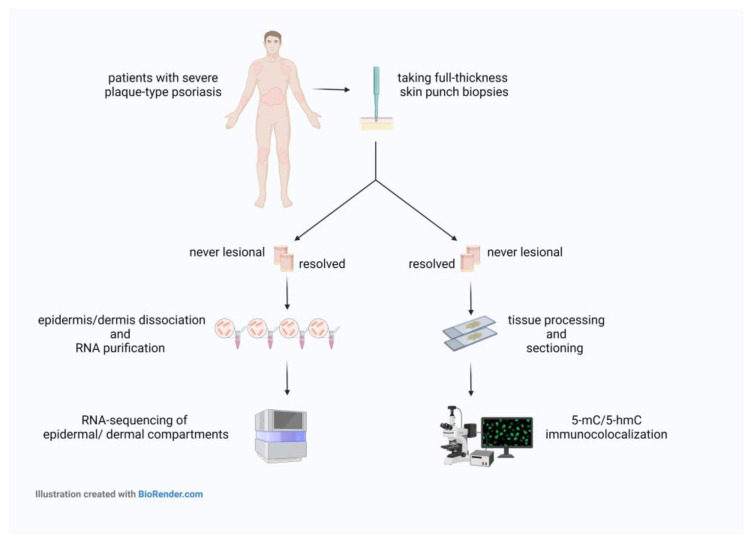
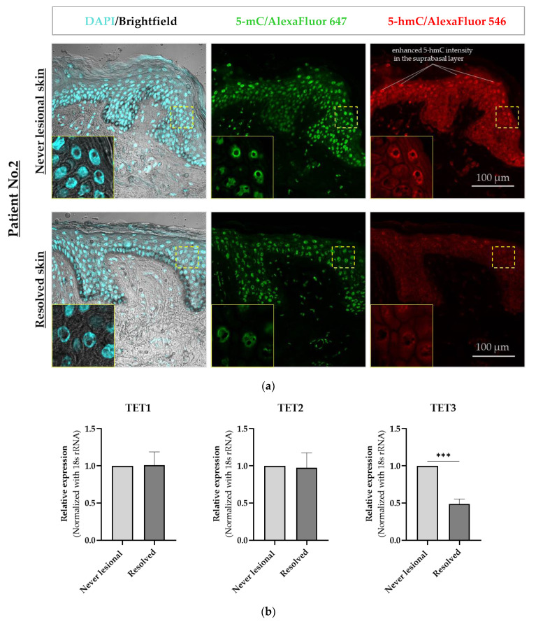
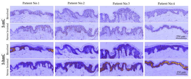
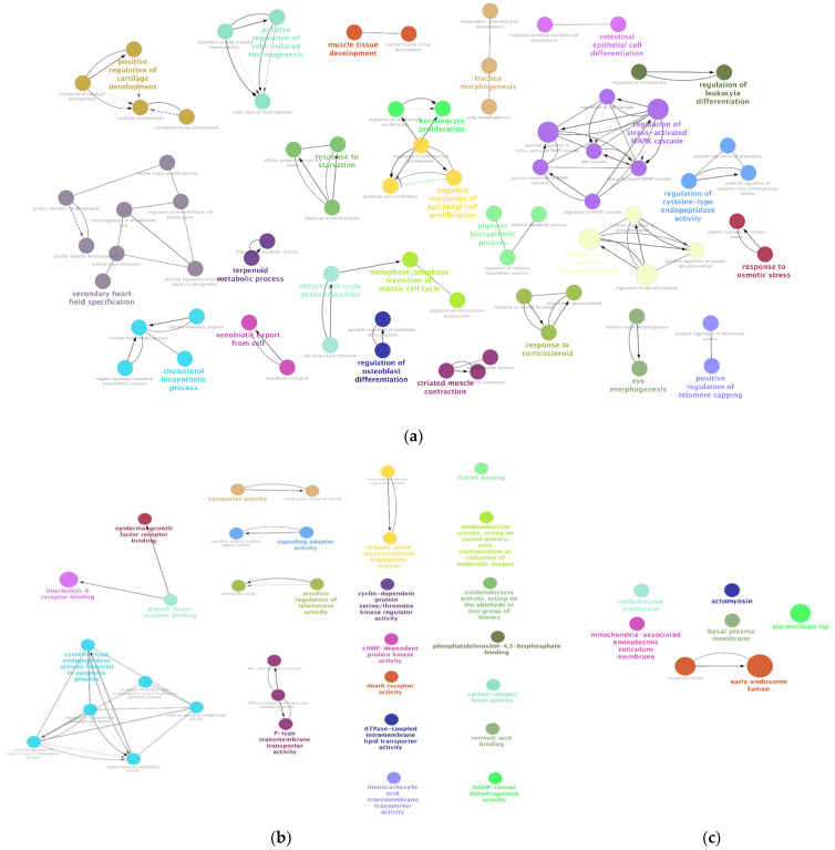
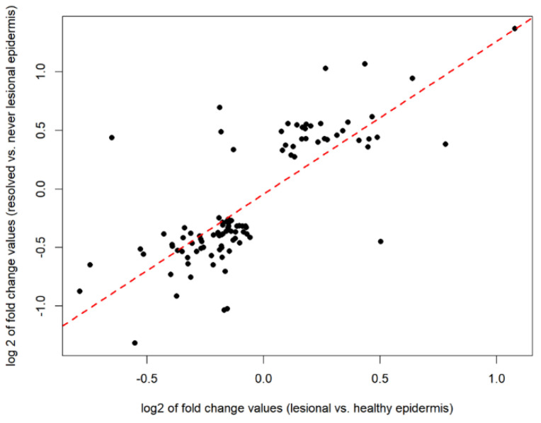
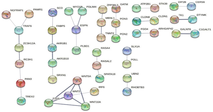
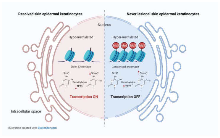
The disease-residual transcriptomic profile (DRTP) within psoriatic healed/resolved skin and epidermal tissue-resident memory T (TRM) cells have been proposed to be crucial for the recurrence of old lesions. However, it is unclear whether epidermal keratinocytes are involved in disease recurrence. There is increasing evidence regarding the importance of epigenetic mechanisms in the pathogenesis of psoriasis. Nonetheless, the epigenetic changes that contribute to the recurrence of psoriasis remain unknown. The aim of this study was to elucidate the role of keratinocytes in psoriasis relapse. The epigenetic marks 5-methylcytosine (5-mC) and 5-hydroxymethylcytosine (5-hmC) were visualized using immunofluorescence staining, and RNA sequencing was performed on paired never-lesional and resolved epidermal and dermal compartments of skin from psoriasis patients. We observed diminished 5-mC and 5-hmC amounts and decreased mRNA expression of the ten-eleven translocation (TET) 3 enzyme in the resolved epidermis. SAMHD1, C10orf99, and AKR1B10: the highly dysregulated genes in resolved epidermis are known to be associated with pathogenesis of psoriasis, and the DRTP was enriched in WNT, TNF, and mTOR signaling pathways. Our results suggest that epigenetic changes detected in epidermal keratinocytes of resolved skin may be responsible for the DRTP in the same regions. Thus, the DRTP of keratinocytes may contribute to site-specific local relapse.

Keywords: 5-hmC; 5-mC; epigenomics; keratinocyte; psoriasis; relapse; transcriptomics.


Conflict of interest statement

The authors declare no conflict of interest.

Figures

Figure 1
Figure 1
Schematic view of the experimental workflow. Abbreviations: 5-mC, 5-methylcytosine; 5-hmC, 5-hydroxymethylcytosine.
Figure 2
Figure 2
Confocal microscopy immunofluorescence analysis of 5-mC and 5-hmC in never-lesional vs. resolved skin. (a) Representative images are shown (n = 4). Colocalization immunofluorescence staining of 5-mC (green) and 5-hmC (red). Closeup images show yellow-dotted regions at higher magnification. Scale bars are 100 μm. (b) Real-time RT-PCR of TET1, TET2, and TET3 in resolved epidermis vs. never-lesional epidermis. An unpaired two-tailed t-test was performed, *** p < 0.001. Data = mean ± SEM of biological replicates (n = 3). Abbreviations: 5-mC, 5-methylcytosine; 5-hmC, 5-hydroxymethylcytosine; TET1, ten-eleven translocation (TET)1; TET2, ten-eleven translocation (TET)2; TET3, ten-eleven translocation (TET)3.
Figure 3
Figure 3
False-colored (pseudocolored) images of skin sections with a low magnification (20×) objective. All never-lesional and resolved pairs are identically brightness-adjusted. Scale bars are 250 μm. Abbreviations: 5-mC, 5-methylcytosine; 5-hmC, 5-hydroxymethylcytosine.
Figure 4
Figure 4
GO enrichment analysis of resolved vs. never-lesional epidermis DEGs. Enrichment shows only significant (a) BPs, (b) MFs, and (c) CCs with p < 0.05. Node color represents the specific functional class, biological processes, and cellular components involved in the enrichment analysis of the identified DEGs. Bold fonts indicate the major BPs, MFs, or CCs that define the names of each group. Abbreviations: DEG, differentially expressed genes; BPs, biological processes; MFs, molecular functions; CCs, cellular components.
Figure 5
Figure 5
The Spearman’s rank correlation coefficient analysis was performed on 102 overlapping genes between 476 DEGs of the resolved vs. never-lesional and the 4104 DEGs of the lesional vs. healthy in epidermal compartments. The amount of change strongly correlated. Abbreviations: DEGs, differentially expressed genes.
Figure 6
Figure 6
PPI networks of the 102 overlapping genes between resolved vs. never-lesional DEGs and lesional vs. healthy DEGs were generated using the STRING database. The figure shows the result ordered by network size. Abbreviations: PPI, protein–protein interaction; DEGs, differentially expressed genes.
Figure 7
Figure 7
Mechanism of action of the three DEGs in resolved vs. never-lesional epidermis identified as likely to be important for lesion development in resolved epidermis. Significantly lower levels of the SAMHD1 gene in resolved epidermal keratinocytes might be accompanied by abnormal dNTP accumulation, which can induce DNA damage. It is established that damaged DNA species can trigger type-I IFN and innate immune responses. At the same time, SAMHD1 higher expression level in never-lesional epidermal keratinocytes may keep DNA integrity more efficiently. Strong up-regulation of GPR15L in resolved epidermal keratinocytes may lead to the recruitment of GPR15+ cells, such as T-cells, into the epidermal compartment. In resolved epidermal keratinocytes, the strong up-regulation of AKR1B10 gene may result in reduced RA and consequently increased keratinocytes survival and proliferation. However, the lower levels of AKR1B10 gene in never-lesional epidermal keratinocytes can activate the retinal–retinoic acid pathway and induce keratinocyte differentiation, while decreasing cell survival. Abbreviations: DEGs, differentially expressed genes; CoA, Coenzyme A; NADPH, nicotinamide adenine dinucleotide phosphate; dNTP, deoxynucleotide triphosphates; PPP, triphosphate; dN, deoxyribonucleoside.
Figure 8
Figure 8
The DNA methylation/hydroxymethylation status of resolved vs. never-lesional epidermal keratinocytes. The DNA was hypo-methylated and hypo-hydroxymethylated in resolved vs. never-lesional epidermal keratinocytes. Hypo-methylated DNA is generally associated with open chromatin and transcription activation. Hence, the transcription might be more active in hypo-methylated DNA of resolved epidermal keratinocytes than never-lesional ones. Furthermore, hypo-methylated DNA in the resolved epidermis can reduce 5-hmC DNA levels. At the same time, the lower mRNA expression of TET3 enzyme might also be another reason for the loss of 5-hmC in the resolved epidermis. Abbreviations: 5-mC, 5-methylcytosine; 5-hmC, 5-hydroxymethylcytosine; TET3, ten-eleven translocation (TET)3.

References

    1. Billi A.C., Gudjonsson J.E., Voorhees J.J. Psoriasis: Past, Present, and Future. J. Investig. Dermatol. 2019;139:e133–e142. doi: 10.1016/j.jid.2019.08.437. - DOI - PMC - PubMed
    1. Owczarek W., Dzik M., Narbutt J., Walecka I., Kowalczyk M. Real-world evidence on time to relapse of plaque psoriasis after discontinuation of biologic treatment in Poland. Dermatol. Ther. 2021;34:e15052. doi: 10.1111/dth.15052. - DOI - PubMed
    1. Bu J., Ding R., Zhou L., Chen X., Shen E. Epidemiology of Psoriasis and Comorbid Diseases: A Narrative Review. Front. Immunol. 2022;13:880201. doi: 10.3389/fimmu.2022.880201. - DOI - PMC - PubMed
    1. Chamian F., Lowes M.A., Lin S.-L., Lee E., Kikuchi T., Gilleaudeau P., Sullivan-Whalen M., Cardinale I., Khatcherian A., Novitskaya I., et al. Alefacept reduces infiltrating T cells, activated dendritic cells, and inflammatory genes in psoriasis vulgaris. Proc. Natl. Acad. Sci. USA. 2005;102:2075–2080. doi: 10.1073/pnas.0409569102. - DOI - PMC - PubMed
    1. Zaba L.C., Cardinale I., Gilleaudeau P., Sullivan-Whalen M., Suárez-Fariñas M., Suárez Fariñas M., Fuentes-Duculan J., Novitskaya I., Khatcherian A., Bluth M.J., et al. Amelioration of epidermal hyperplasia by TNF inhibition is associated with reduced Th17 responses. J. Exp. Med. 2007;204:3183–3194. doi: 10.1084/jem.20071094. - DOI - PMC - PubMed