Review
. 2023 Feb 26;24(5):4580.
doi: 10.3390/ijms24054580.

Pyodermitis during Nivolumab Treatment for Non-Small Cell Lung Cancer: A Case Report and Review of the Literature

Affiliations
Review

Pyodermitis during Nivolumab Treatment for Non-Small Cell Lung Cancer: A Case Report and Review of the Literature

Terenzio Cosio et al. Int J Mol Sci. .

Abstract

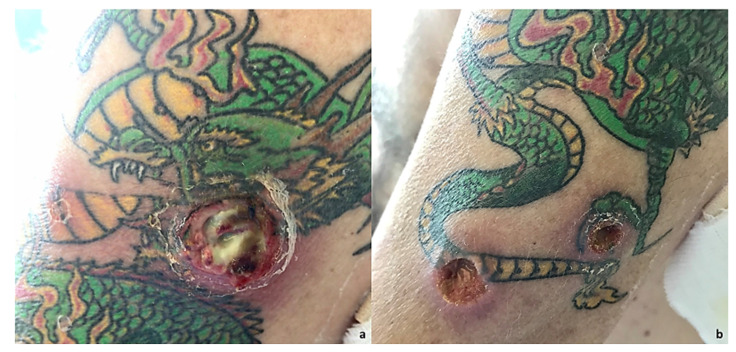
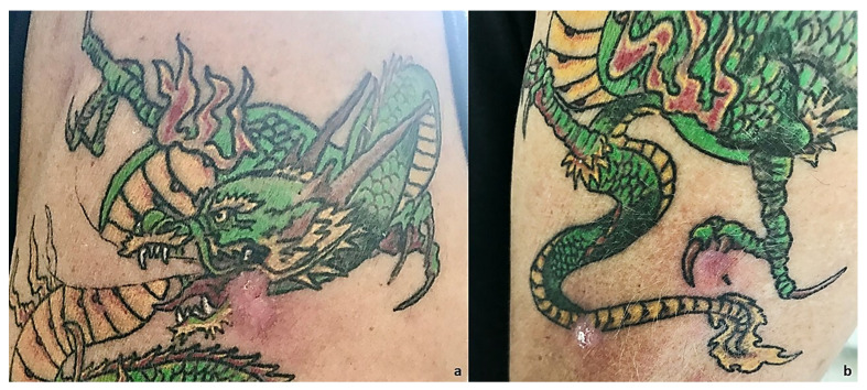
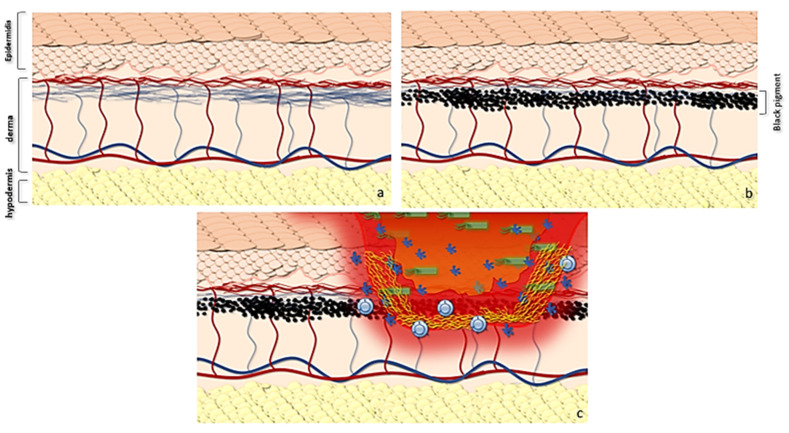
Immunotherapy in oncology is replacing traditional therapies due to it specific action and limited side effects. Despite the high efficacy of immunotherapy, side effects such as bacterial infection have been reported. Bacterial skin and soft tissue infections represent one of the most important differential diagnoses in patients presenting with reddened and swollen skin and soft tissue. Among these infections, cellulitis (phlegmon) and abscesses are the most frequent. In most cases, these infections occur locally with possible contiguous spread, or as a multifocal manifestation, especially in immunocompromised patients. Herein, we report a case of pyodermitis in an immunocompromised district in a patient treated with nivolumab for non-small cell lung cancer. A 64-year-old, smoker male patient showed cutaneous lesions at a different evolution level in the left arm, all in a tattooed area, with one phlegmon and two ulcerated lesions. Microbiological cultures and gram staining revealed an infection caused by a methicillin-susceptible but erythromycin-resistant (ER-R), clindamycin-resistant (CL-R), and gentamicin-resistant (GE-R) Staphylococcus aureus strain. Despite immunotherapy becoming a milestone in oncologic treatment, more than the spectrum of immune-mediated toxicities of these agents needs to be investigated. This report highlights the importance of considering lifestyle and cutaneous background before starting immunotherapy for cancer treatment, with an emphasis on pharmacogenomics and the possibility of modified skin microbiota predisposing to cutaneous infections in patients treated with PD-1 inhibitors.

Keywords: Staphylococcus aureus; cutaneous infection; immunotherapy; nivolumab.


Conflict of interest statement

The authors declare no conflict of interest.

Figures

Figure 1
Figure 1
The therapy response timeline from 2015 to today. (a) Chest X-ray (Thoravision). Examination performed with digital technologies in the two orthogonal projections. The parenchymal opacity already indicated on the right and better evident in the previous TC of 3/3/2015 is confirmed. Disventilatory streak in the left basal site. (b) Examination performed before and after intravenous administration of organ-iodized contrast medium place comparison with the previous PET-CT investigation carried out on 29 July 2016. Compared to the previous one, there is a documented increase in the size of the newly formed tissue located in the right hilar seat (current dimensions 27 × 25 mm vs. 20 × 11 mm of the previous exam). The findings in the lungs are unchanged, the thickening area previously described at the level of the LID. No pleural effusion flaps. (c) Examination carried out before and after administration of organ-iodized MDC by IV route, compared with previous PET-CT carried out on 5 July 2018. There are no areas of pathological enhancement in the sub- and supratentorial sites. The axial ventricular system showed dimensions within the limits of the norm. The results of the surgery known in the anamnesis in the thoracic area on the right have been confirmed. The millimetric nodular formation already evident at the lower lobe appears unchanged right in the previous aforementioned signs of emphysema in both parenchymal areas. No pleural effusion flaps have been observed. (d) Neck-thorax: Nodular thyroid isthmic formation requiring monitoring remains unchanged instrumental with ultrasound examination. The results of surgery on the upper-right lobe have been confirmed. The share of tissue in the right hilar seat and the solid nodular formation (4 mm) correspond to the LID. No pleural or pericardial effusion. Sub-centimetric lymph node formations in the hilar and mediastinal region were stable in size.
Figure 2
Figure 2
(a) At an objective examination, it is possible to notice an exudative lesion of the skin with a violet halo index of subcutaneous tissue infection compatible with a suppurative bacterial infection. The patient stated he had pain on the inspection site. Moreover, pain, tumor, calor, rubror, all signs of acute inflammation, were detectable. Clinical diagnosis of phlegmon was made. (b) On physical examination, it is possible to notice two lesions arising in the tattooed area. The lesions are excavated, with a clean margin and a fibrous bottom, where it is possible to see the onset of skin infection; then, infection is confirmed through microbiological tests. Clinical signs of subcutaneous tissue inflammation, i.e., pain, tumor, and perilesional rubror were detectable. Circular areas of superficial flaking, as reported by the patient, also occurred as primary lesions followed by skin ulceration.
Figure 3
Figure 3
(a) Clinical resolution of the phlegmon after 15 days. (b) Clinical resolution with topical fusidic acid in combination with oral clarithromycin 250 mg/daily for 15 days.
Figure 4
Figure 4
Graphic abstract showing the possible evolution of gram+ skin infections in tattooed patients with immunotherapy. (a) Normal skin anatomy and trophism of epidermis. (b) The use of the pigment leads both to a local microtrauma and to the deposit of pigment in the superficial portion of the dermis with processes of inflammation at the time of tattooing and subsequent inflammation. At the same time, the pigment, together with microtrauma fibrosis, reduces the diffusion of transudate from the dermis to the epidermis, also decreasing the superficial trophism. (c) This determines a reduced barrier action of the epidermis, which supports Ruocco’s hypothesis. In LMR, infections can lead to a local destructive process evolving in phlegmon.

Similar articles

Cited by

References

    1. Topalian S.L., Hodi F.S., Brahmer J.R., Gettinger S.N., Smith D.C., McDermott D.F., Powderly J.D., Carvajal R.D., Sosman J.A., Atkins M.B., et al. Safety, Activity, and Immune Correlates of Anti-PD-1 Antibody in Cancer. N. Engl. J. Med. 2012;366:2443–2454. doi: 10.1056/NEJMoa1200690. - DOI - PMC - PubMed
    1. Lu T., Yang X., Huang Y., Zhao M., Li M., Ma K., Yin J., Zhan C., Wang Q. Trends in the incidence, treatment, and survival of patients with lung cancer in the last four decades. Cancer Manag. Res. 2019;11:943–953. doi: 10.2147/CMAR.S187317. - DOI - PMC - PubMed
    1. Lim S.M., Hong M.H., Kim H.R. Immunotherapy for Non-small Cell Lung Cancer: Current Landscape and Future Perspectives. Immune Netw. 2020;20:e10. doi: 10.4110/in.2020.20.e10. - DOI - PMC - PubMed
    1. Dang T.O., Ogunniyi A., Barbee M.S., Drilon A. Pembrolizumab for the treatment of PD-L1 positive advanced or metastatic non-small cell lung cancer. Expert Rev. Anticancer. Ther. 2016;16:13–20. doi: 10.1586/14737140.2016.1123626. - DOI - PMC - PubMed
    1. Brahmer J.R., Govindan R., Anders R.A., Antonia S.J., Sagorsky S., Davies M.J., Dubinett S.M., Ferris A., Gandhi L., Garon E.B., et al. The Society for Immunotherapy of Cancer consensus statement on immunotherapy for the treatment of non-small cell lung cancer (NSCLC) J. Immunother. Cancer. 2018;6:75. doi: 10.1186/s40425-018-0382-2. - DOI - PMC - PubMed