. 2023 Feb 26;24(5):4585.
doi: 10.3390/ijms24054585.

Low Dose of BPA Induces Liver Injury through Oxidative Stress, Inflammation and Apoptosis in Long-Evans Lactating Rats and Its Perinatal Effect on Female PND6 Offspring

Affiliations

Low Dose of BPA Induces Liver Injury through Oxidative Stress, Inflammation and Apoptosis in Long-Evans Lactating Rats and Its Perinatal Effect on Female PND6 Offspring

Beatriz Linillos-Pradillo et al. Int J Mol Sci. .

Abstract

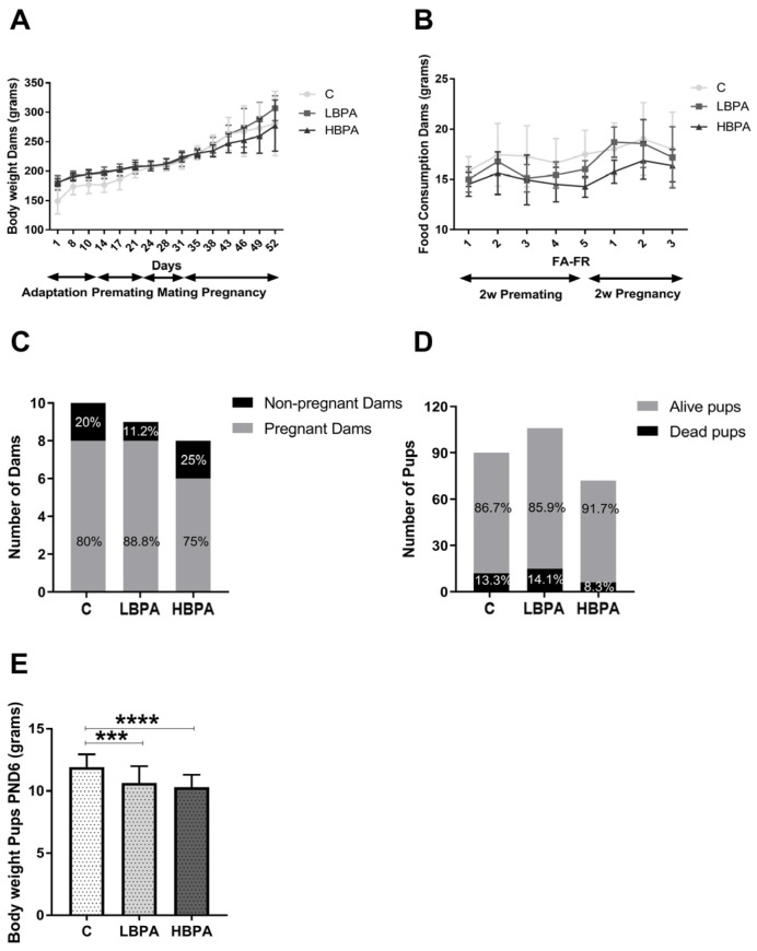
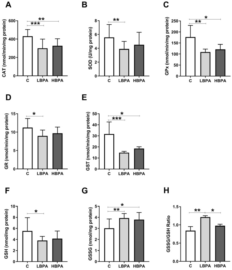
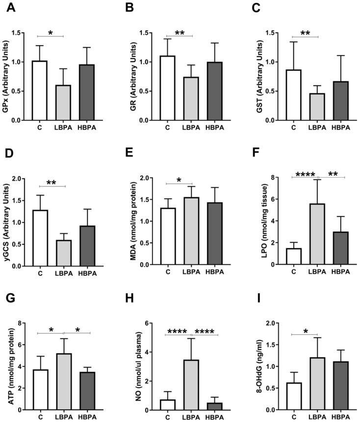
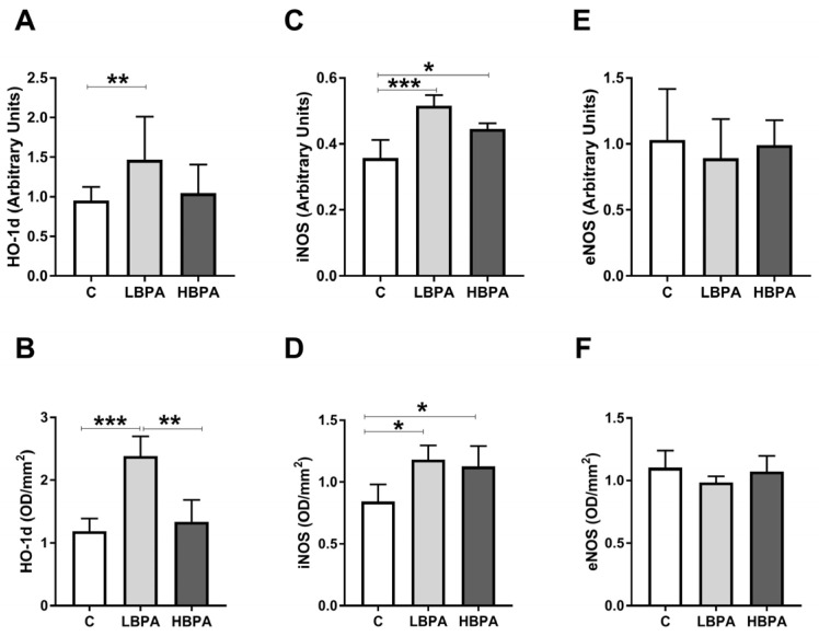
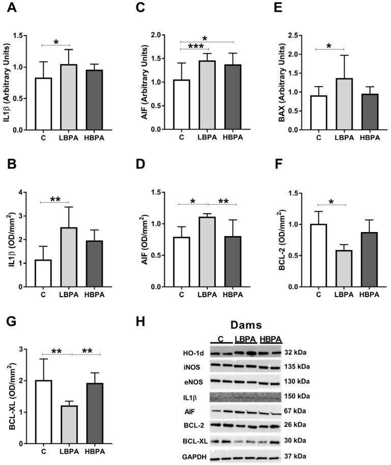
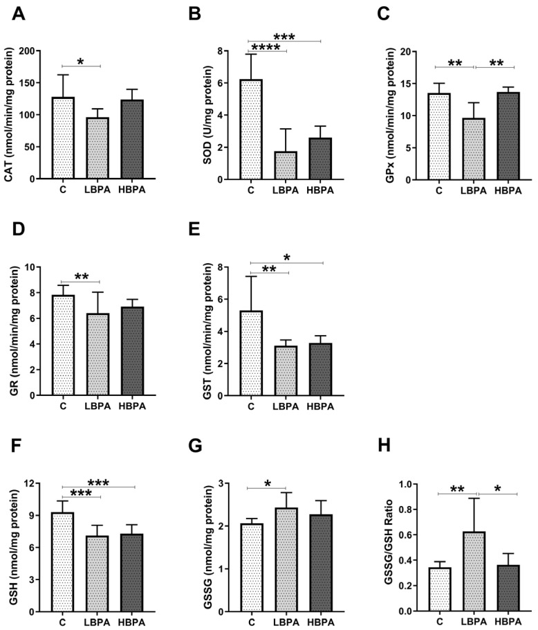
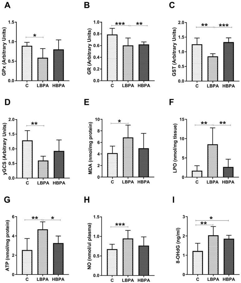
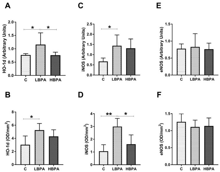
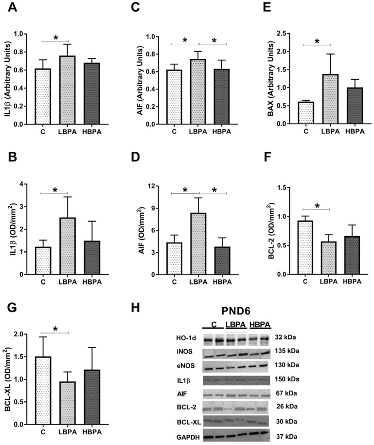
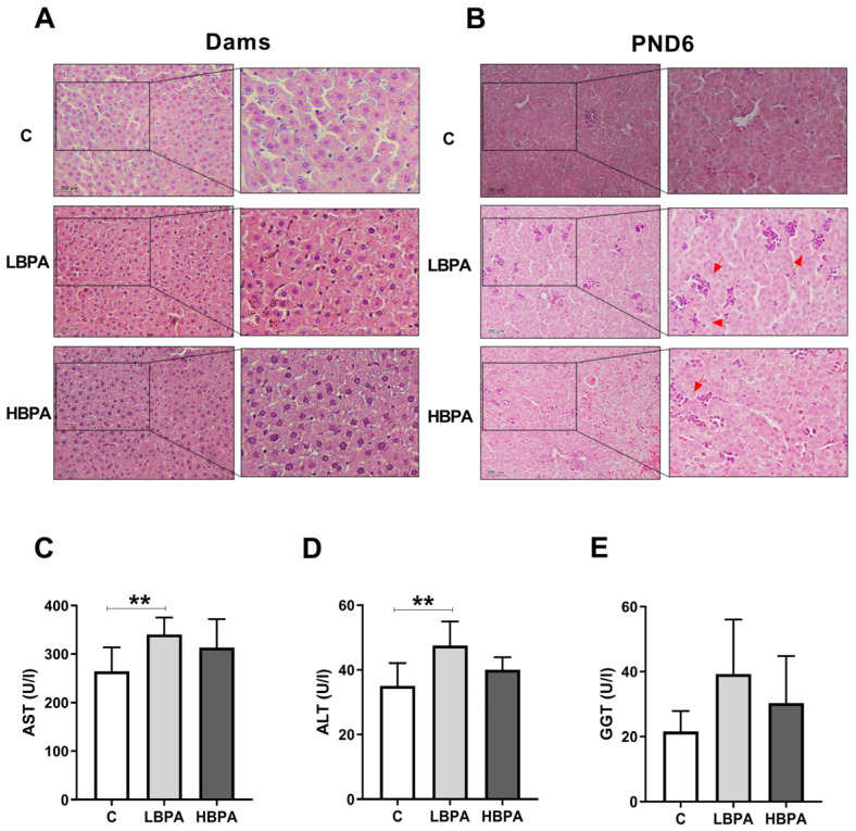
Bisphenol A (BPA) is a phenolic compound used in plastics elaboration for food protection or packaging. BPA-monomers can be released into the food chain, resulting in continuous and ubiquitous low-dose human exposure. This exposure during prenatal development is especially critical and could lead to alterations in ontogeny of tissues increasing the risk of developing diseases in adulthood. The aim was to evaluate whether BPA administration (0.036 mg/kg b.w./day and 3.42 mg/kg b.w./day) to pregnant rats could induce liver injury by generating oxidative stress, inflammation and apoptosis, and whether these effects may be observed in female postnatal day-6 (PND6) offspring. Antioxidant enzymes (CAT, SOD, GR, GPx and GST), glutathione system (GSH/GSSG) and lipid-DNA damage markers (MDA, LPO, NO, 8-OHdG) were measured using colorimetric methods. Inducers of oxidative stress (HO-1d, iNOS, eNOS), inflammation (IL-1β) and apoptosis (AIF, BAX, Bcl-2 and BCL-XL) were measured by qRT-PCR and Western blotting in liver of lactating dams and offspring. Hepatic serum markers and histology were performed. Low dose of BPA caused liver injury in lactating dams and had a perinatal effect in female PND6 offspring by increasing oxidative stress levels, triggering an inflammatory response and apoptosis pathways in the organ responsible for detoxification of this endocrine disruptor.

Keywords: apoptosis; bisphenol A; inflammation; liver injury; oxidative stress; perinatal offspring.


Conflict of interest statement

The authors declare no conflict of interest.

Figures

Figure 1
Figure 1
Body weight, food consumption and reproduction data in pregnant dams and neonatal offspring. (A) Body weight of dams monitored every 3–4 days; (B) food consumption during the second week of premating and the second week of pregnancy; (C) number of pregnant and non-pregnant dams; (D) percentage of alive and dead offspring after birth and (E) body weight of PND6 offspring after birth. Data are represented as mean ± SD. Statistical significance was determined by one-way ANOVA. *** p < 0.001, **** p < 0.0001.
Figure 2
Figure 2
Antioxidant enzyme activities and glutathione concentrations in livers from lactating dams after exposure to low and high doses of BPA. (A) Enzymatic activity of catalase (CAT) in nmol/min/mg protein; (B) superoxide dismutase (SOD) in U/mg protein; (C) glutathione peroxidase (GPx) in nmol/min/mg protein; (D) glutathione reductase (GR) in nmol/min/mg protein; (E) glutathione S-transferase (GST) in nmol/min/mg protein. (F) Concentration of reduced glutathione (GSH) in nmol/mg protein; (G) concentration of oxidized glutathione (GSSG) in nmol/mg protein. (H) GSSG/GSH ratio. Data represent mean ± SD. n = 8 control dams; n = 8 LBPA dams; n = 6 HBPA dams (two replicates for each sample). Statistical significance was determined by one-way ANOVA. * p < 0.05; ** p < 0.01; *** p < 0.001.
Figure 3
Figure 3
Gene expression profile of GSH-related enzymes and parameters of oxidative damage in livers from lactating dams after exposure to different doses of BPA. (A) Glutathione peroxidase (GPx), (B) glutathione reductase (GR), (C) glutathione S-transferase (GST) and (D) γ-glutamylcysteine synthetase (γ-GCS) relative gene expression. (E) Malondialdehyde (MDA) content in nmol/mg protein. (F) Lipid hydroperoxide (LPO) content in nmol/mg tissue. (G) Adenosine triphosphate (ATP) levels in nmol/mg protein. (H) Concentration of nitric oxide metabolites (NO) in nmol/μL plasma. (I) Concentration of 8-oxo-2′-deoxyguanosine (8-OHdG) in ng/mL. Data represent mean ± SD. For mRNA, n = 8 control dams; n = 8 LBPA dams; n = 6 HBPA dams (three replicates for each gene). For ELISA assay, n = 8 control dams; n = 8 LBPA mg/kg dams; n = 6 HBPA dams (two replicates for each sample). Statistical significance was determined by one-way ANOVA. * p < 0.05; ** p < 0.01; **** p < 0.0001.
Figure 4
Figure 4
Oxidative stress intermediaries in livers from lactating dams after exposure to different doses of BPA. mRNA and protein expressions of HO-1d (heme oxygenase 1) (A,B); iNOS (inducible nitric oxide synthase) (C,D); and eNOS (endothelial nitric oxide synthase) (E,F). Data represent mean ± SD. For mRNA, n = 8 control dams; n = 8 LBPA dams; n = 6 HBPA dams (three replicates for each gene). For protein, n = 5 rats per experimental group. Data represent mean ± SD. Statistical significance was determined by one-way ANOVA. * p < 0.05; ** p < 0.01; *** p < 0.001.
Figure 5
Figure 5
Inflammatory mediator and apoptosis markers in livers from lactating dams after exposure to different doses of BPA. mRNA and protein expressions of IL1β (interleukin-1-β) (A,B); mRNA and protein expression of AIF (apoptosis-inducing factor) (C,D). (E) mRNA expression of BAX (Bcl-2-associated X protein). (F,G) Protein expressions of BCL-2 (B-cell lymphoma) and BCL-XL (B-cell lymphoma-extra large). (H) Representative images of the Western blot results of the different proteins studied. Data represent mean ± SD. For mRNA, n = 8 control dams; n = 8 LBPA dams; n = 6 HBPA dams (three replicates for each gene). For protein, n = 5 rats per experimental group. Statistical significance was determined by one-way ANOVA. * p < 0.05; ** p < 0.01; *** p < 0.001.
Figure 6
Figure 6
Effects of perinatal exposure to BPA on antioxidant enzyme activities and glutathione concentrations in livers from female PND6 offspring. (A) Enzymatic activity of catalase (CAT) in nmol/min/mg protein; (B) superoxide dismutase (SOD) in U/mg protein; (C) glutathione peroxidase (GPx) in nmol/min/mg protein; (D) glutathione reductase (GR) in nmol/min/mg protein; (E) glutathione S-transferase (GST) in nmol/min/mg protein. (F) Concentration of reduced glutathione (GSH) in nmol/mg protein; (G) concentration of oxidized glutathione (GSSG) in ng/mg protein. (H) GSSG/GSH ratio. Data represent mean ± SD. n = 12 control PND6 offspring; n = 12 LBPA PND6 offspring; n = 12 HBPA PND6 offspring (two replicates for each sample). Statistical significance was determined by one-way ANOVA. * p < 0.05; ** p < 0.01; *** p < 0.001; **** p < 0.0001.
Figure 7
Figure 7
Effects of perinatal exposure to BPA on gene expression profile of GSH-related enzymes and oxidative damage in livers from female PND6 offspring. (A) Glutathione peroxidase (GPx); (B) glutathione reductase (GR); (C) glutathione S-transferase (GST); (D) γ-glutamylcysteine synthetase (γ-GCS) relative gene expression. (E) Malondialdehyde (MDA) content in nmol/mg protein. (F) Lipid hydroperoxide content in nmol/mg tissue. (G) Adenosine triphosphate (ATP) levels in nmol/mg protein. (H) Concentration of nitric oxide metabolites (NO) in nmol/μL plasma. (I) Concentration of 8-oxo-2′-deoxyguanosine (8-OHdG) in ng/mL. Data represent mean ± SD. For ELISA assay, n = 12 control PND6 offspring; n = 12 LBPA PND6 offspring; n = 12 HBPA PND6 offspring (two replicates for each sample). For mRNA, n = 12 control PND6 offspring; n = 12 LBPA PND6 offspring; n = 12 HBPA PND6 offspring (three replicates for each gene). Statistical significance was determined by one-way ANOVA. * p < 0.05; ** p < 0.01; *** p < 0.001.
Figure 8
Figure 8
Effects of perinatal exposure to BPA on oxidative stress intermediaries in livers from female PND6 offspring. mRNA and protein expressions of HO-1d (heme oxygenase 1) (A,B); iNOS (inducible nitric oxide synthase) (C,D); and eNOS (endothelial nitric oxide synthase) (E,F). Data represent mean ± SD. For mRNA, n = 12 control PND6 offspring; n = 12 LBPA PND6 offspring; n = 12 HBPA PND6 offspring (three replicates for each gene). For protein, n = 5 rats per experimental group. Statistical significance was determined by one-way ANOVA. * p < 0.05; ** p < 0.01.
Figure 9
Figure 9
Effects of perinatal exposure to BPA on inflammatory mediator and apoptosis markers in livers from female PND6 offspring. (A,B) mRNA and protein expressions of IL1β (interleukin-1-β). (C,D); mRNA and protein expression of AIF (apoptosis-inducing factor). (E) mRNA of BAX (Bcl-2-associated X protein). (F,G) Protein expressions of BCL-2 (B-cell lymphoma) and BCL-XL (B-cell lymphoma-extra large). (H) Representative images of the Western blot results of the different proteins studied. Data represent mean ± SD. For mRNA, n = 12 control PND6 offspring; n = 12 LBPA PND6 offspring; n = 12 HBPA PND6 offspring (three replicates for each gene). For protein, n = 5 rats per experimental group. Statistical significance was determined by one-way ANOVA. * p < 0.05.
Figure 10
Figure 10
Histology and clinical chemistry of liver functional markers after BPA administration. Representative images of livers from dams (A) and female PND6 offspring (B) stained with H&E in control and low and high BPA treatment groups (LBPA or HBPA). Scale bars indicate 200 µm (20×) and magnified image of the characteristic tissue section (40×). Red arrows indicate the aggregation of nuclei. (C) Aspartate aminotransferase (AST); (D) alanine aminotransferase (ALP) and (E) gamma glutamyl transpeptidase (GGT) in U/L in serum dams. Data represent mean ± SD. n = 8 control dams; n = 8 LBPA dams; n = 6 HBPA dams. Statistical significance was determined by one-way ANOVA. ** p < 0.01.
Figure 11
Figure 11
Experimental design. The diet of the parental generation (F0) was different according to the experimental group (control, BPA 0.036 mg/kg and BPA 3.42 mg/kg). During the entire experiment (premating, mating, pregnancy, lactation), the treatment was maintained until the time of dissection in dams and pups to postnatal day 6 (PND6). The influence of lactating dams and perinatal exposure to BPA on livers and its possible mechanism in offspring were studied. Figure created with Prism v7 (GraphPad Software Inc., La Jolla, CA, USA).

Similar articles

Cited by

References

    1. Kazemi S., Mousavi S.N., Aghapour F., Rezaee B., Sadeghi F., Moghadamnia A.A. Induction Effect of Bisphenol a on Gene Expression Involving Hepatic Oxidative Stress in Rat. Oxid. Med. Cell. Longev. 2016;2016:12–14. doi: 10.1155/2016/6298515. - DOI - PMC - PubMed
    1. Zhang Y., Shi Y., Li Z., Sun L., Zhang M., Yu L., Wu S. BPA Disrupts 17-estradiol-mediated Hepatic Protection against Ischemia/Reperfusion Injury in Rat Liver by Upregulating the Ang II/AT1R Signaling Pathway. Mol. Med. Rep. 2020;22:416–422. doi: 10.3892/mmr.2020.11072. - DOI - PMC - PubMed
    1. Acaroz U., Ince S., Arslan-Acaroz D., Gurler Z., Demirel H.H., Kucukkurt I., Eryavuz A., Kara R., Varol N., Zhu K. Bisphenol-A Induced Oxidative Stress, Inflammatory Gene Expression, and Metabolic and Histopathological Changes in Male Wistar Albino Rats: Protective Role of Boron. Toxicol. Res. 2019;8:262–269. doi: 10.1039/C8TX00312B. - DOI - PMC - PubMed
    1. Peerapanyasut W., Kobroob A., Palee S., Chattipakorn N., Wongmekiat O. Activation of Sirtuin 3 and Maintenance of Mitochondrial Integrity by N-Acetylcysteine Protects against Bisphenol A-Induced Kidney and Liver Toxicity in Rats. Int. J. Mol. Sci. 2019;20:267. doi: 10.3390/ijms20020267. - DOI - PMC - PubMed
    1. Eweda S., Newairy A., Abdou H., Gaber A. Bisphenol A-induced Oxidative Damage in the Hepatic and Cardiac Tissues of Rats: The Modulatory Role of Sesame Lignans. Exp. Ther. Med. 2019;2:33–44. doi: 10.3892/etm.2019.8193. - DOI - PMC - PubMed