Review
. 2023 Mar 1;24(5):4746.
doi: 10.3390/ijms24054746.

When Just One Phosphate Is One Too Many: The Multifaceted Interplay between Myc and Kinases

Affiliations
Review

When Just One Phosphate Is One Too Many: The Multifaceted Interplay between Myc and Kinases

Dalila Boi et al. Int J Mol Sci. .

Abstract

Myc transcription factors are key regulators of many cellular processes, with Myc target genes crucially implicated in the management of cell proliferation and stem pluripotency, energy metabolism, protein synthesis, angiogenesis, DNA damage response, and apoptosis. Given the wide involvement of Myc in cellular dynamics, it is not surprising that its overexpression is frequently associated with cancer. Noteworthy, in cancer cells where high Myc levels are maintained, the overexpression of Myc-associated kinases is often observed and required to foster tumour cells' proliferation. A mutual interplay exists between Myc and kinases: the latter, which are Myc transcriptional targets, phosphorylate Myc, allowing its transcriptional activity, highlighting a clear regulatory loop. At the protein level, Myc activity and turnover is also tightly regulated by kinases, with a finely tuned balance between translation and rapid protein degradation. In this perspective, we focus on the cross-regulation of Myc and its associated protein kinases underlying similar and redundant mechanisms of regulation at different levels, from transcriptional to post-translational events. Furthermore, a review of the indirect effects of known kinase inhibitors on Myc provides an opportunity to identify alternative and combined therapeutic approaches for cancer treatment.

Keywords: Aurora-A; Aurora-B; BRD4; GSK-3; Myc; PIM; PKA; PLK1; kinase inhibitors; kinases.


Conflict of interest statement

The authors declare no conflict of interest.

Figures

Figure 1
Figure 1
Activation and phospho-dependent stabilization of Myc. (A) Myc can be activated in response to different stimuli via several transduction pathways converging to Myc stabilization. Stabilized Myc associates with its protein partner MAX to form a heterodimer, which, together with other co-activators, binds to the E-box elements and drives the transcription of a target gene subset involved in a wide range of cellular processes. (B) Phospho-dependent stabilization of Myc. Myc displays two key phosphorylation sites that undergo hierarchical phosphorylation, supervising protein stability. Phosphorylation of Myc at the S62 residue determines protein stabilization and primes the subsequent phosphorylation at T58, which induces the removal of the phosphate group at S62; the unstable, singly phosphorylated T58-Myc is then recognized by the ubiquitin ligase Fbxw7 and degraded by the ubiquitin–proteasome system. Created with BioRender.com (accessed on 20 January 2023).
Figure 1
Figure 1
Activation and phospho-dependent stabilization of Myc. (A) Myc can be activated in response to different stimuli via several transduction pathways converging to Myc stabilization. Stabilized Myc associates with its protein partner MAX to form a heterodimer, which, together with other co-activators, binds to the E-box elements and drives the transcription of a target gene subset involved in a wide range of cellular processes. (B) Phospho-dependent stabilization of Myc. Myc displays two key phosphorylation sites that undergo hierarchical phosphorylation, supervising protein stability. Phosphorylation of Myc at the S62 residue determines protein stabilization and primes the subsequent phosphorylation at T58, which induces the removal of the phosphate group at S62; the unstable, singly phosphorylated T58-Myc is then recognized by the ubiquitin ligase Fbxw7 and degraded by the ubiquitin–proteasome system. Created with BioRender.com (accessed on 20 January 2023).
Figure 2
Figure 2
Various mechanisms by which PLK1 stabilizes Myc. PLK1 can directly phosphorylate Myc at S62 or indirectly promote phosphorylation at S281 in a PKA-dependent manner, resulting in the accumulation of a stable, ubiquitinylated form of Myc through β-TrCP binding (right side). Unlike Fbxw7, in fact, β-TrCP assembles K33/K63/K48 heterotypic polyubiquitin chains that do not target Myc for proteasome-mediated degradation. PLK1 also impairs Myc degradation by promoting the proteolysis of Fbxw7, thereby increasing the half-life of Myc. Stabilized Myc, in turn, enhances PLK1 transcription. These mechanisms work together to help maintain high levels of Myc in MYC-amplified tumours, which is associated with poor prognosis. The inhibitors acting on this pathway are shown (see also Section 5.1 for references). Created with BioRender.com (accessed on 20 January 2023).
Figure 3
Figure 3
Schematic exemplification of the main functional interplays between Myc and Aurora kinase A (left)/B(right). Aurora-A physically interacts with N-Myc to form a complex that prevents N-Myc from the proteolytic degradation mediated by the ubiquitin ligase Fbxw7. From a transcriptional point of view, Aurora-A contributes to regulating MYCC expression in combination with hnRNP K. Furthermore, Aurora-A is a target gene of MYCC, which enhances Aurora-A transcription. A similar regulatory relationship is described for MYCN and Aurora-B, with MYCN able to bind to motifs upstream of the Aurora B transcription starting site. A kinase-dependent stabilization of Myc involves phosphorylation at serine residue 67, which is a characteristic feature of Aurora kinase B. The inhibitors acting on this pathway are shown (see also Section 5.2 for references). Created with BioRender.com (accessed on 20 January 2023).
Figure 4
Figure 4
Main Myc regulatory mechanisms by GSK-3. GSK-3 can directly phosphorylate Myc at T58, thus destabilizing the protein and committing it to degradation. In addition, GSK-3 phosphorylates β-catenin and induces its proteolysis, preventing its nuclear translocation. The phosphorylation of GSK-3 at S9/S21 by different kinases prevents both Myc and β-catenin degradation and determines both Myc stabilization, as well as nuclear localization of β-catenin. In the nucleus, β-catenin functions as a coactivator of the TCL/LEF transcription factors and promotes MYC transcription. The main inhibitor acting on this pathway is shown (see also Section 5.3 for references). Created with BioRender.com (accessed on 20 January 2023).
Figure 5
Figure 5
Myc is stabilized via a PKA/PLK1/Myc axis. Myc is stabilized via a PKA/PLK1/Myc axis, which triggers the sequential phosphorylation of the residue S279 by PKA and the residue S281 by PLK1. Stabilized Myc, in turn, enhances the transcription of PKA and, in particular, of the subunit PKA-Cβ, highlighting the relevance of PKA isoform-specific regulatory mechanisms. The main inhibitor acting on this pathway is shown (see also Section 5.4 for references). Created with BioRender.com (accessed on 20 January 2023).
Figure 6
Figure 6
Regulation of Myc by PIM kinases. PIMs phosphorylate Myc at different residues with an isoform specificity and contribute to enhancing protein stability. In addition, PIMs contribute to epigenetic modulation by forming a ternary complex with MYC-MAX, thus regulating the expression of genes under MYC transcriptional control. PIM, in fact, phosphorylates the histone H3 within the E-box element of MYC-dependent genes at the serine residue 10. Phosphorylated H3 recruits the phosphoserine-binding protein 14-3-3, which interacts with the histone acetyltransferase MOF. MOF, in turn, catalyses the acetylation of the histone H4 at K16. Acetylated histone H4 provides a binding platform for BRD4 and p-TEFb, facilitating transcriptional elongation. The inhibitors acting on this pathway are shown (see also Section 5.5 for references). To simplify, dual inhibitors are not reported in the figure. Created with BioRender.com (accessed on 20 January 2023).
Figure 7
Figure 7
BRD4-dependent regulation of Myc. BRD4 directly phosphorylates Myc at T58 through its kinase activity, thus inducing its degradation and reducing its inhibitory effect on BRD4 histone acetyltransferase activity. However, the physical interaction of Myc and BRD4 with ERK1 to form a ternary complex prevents Myc from BRD4-mediated destabilization. Furthermore, BRD4 promotes MYC transcriptional activation by decondensing the chromatin around the MYC gene by means of its histone acetyltransferase activity. The main inhibitors acting on this pathway are shown (see also Section 5.6 for references). To simplify, dual inhibitors are not reported in the figure. Created with BioRender.com (accessed on 20 January 2023).
Figure 8
Figure 8
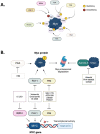
Graphical summary of the kinases and the key phosphorylation sites involved in Myc protein stability are displayed in panel (A). Panel (B) schematizes the principal inhibitors affecting the pathways exploited to indirectly target Myc at both the protein and gene level. Created with BioRender.com (accessed on 20 January 2023).

References

    1. Scafuro M., Capasso L., Carafa V., Altucci L., Nebbioso A. Gene transactivation and transrepression in myc-driven cancers. Int. J. Mol. Sci. 2021;22:3458. doi: 10.3390/ijms22073458. - DOI - PMC - PubMed
    1. Sheiness D., Bishop J.M. DNA and RNA from Uninfected Vertebrate Cells Contain Nucleotide Sequences Related to the Putative Transforming Gene of Avian Myelocytomatosis Virus. J. Virol. 1979;31:514–521. doi: 10.1128/jvi.31.2.514-521.1979. - DOI - PMC - PubMed
    1. Roussel M., Saule S., Lagrou C., Rommens C., Beug H., Graf T., Stehelin D. Three new types of viral oncogene of cellular origin specific for haematopoietic cell transformation. Nature. 1979;281:452–455. doi: 10.1038/281452a0. - DOI - PubMed
    1. Schwab M., Alitalo K., Klempnauer K.-H., Varmus H.E., Bishop J.M., Gilbert F., Brodeur G., Goldstein M., Trent J. Amplified DNA with limited homology to myc cellular oncogene is shared by human neuroblastoma cell lines and a neuroblastoma tumour. Nature. 1983;305:245–248. doi: 10.1038/305245a0. - DOI - PubMed
    1. Kohl N.E., Kanda N., Schreck R.R., Bruns G., Latt S.A., Gilbert F., Alt F.W. Transposition and amplification of oncogene-related sequences in human neuroblastomas. Pt 1Cell. 1983;35:359–367. doi: 10.1016/0092-8674(83)90169-1. - DOI - PubMed

MeSH terms

LinkOut - more resources