. 2023 Mar 2;24(5):4851.
doi: 10.3390/ijms24054851.

Oxidative Status Determines the Cytotoxicity of Ascorbic Acid in Human Oral Normal and Cancer Cells

Affiliations

Oxidative Status Determines the Cytotoxicity of Ascorbic Acid in Human Oral Normal and Cancer Cells

Wei-Zhi Huang et al. Int J Mol Sci. .

Abstract

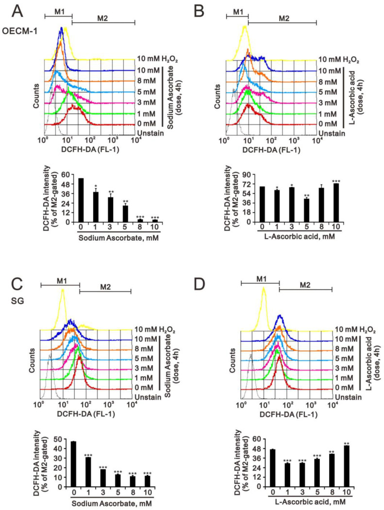
Oral squamous cell carcinoma (OSCC) can arise anywhere in the oral cavity. OSCC's molecular pathogenesis is complex, resulting from a wide range of events that involve the interplay between genetic mutations and altered levels of transcripts, proteins, and metabolites. Platinum-based drugs are the first-line treatment for OSCC; however, severe side-effects and resistance are challenging issues. Thus, there is an urgent clinical need to develop novel and/or combinatory therapeutics. In this study, we investigated the cytotoxic effects of pharmacological concentrations of ascorbate on two human oral cell lines, the oral epidermoid carcinoma meng-1 (OECM-1) cell and the Smulow-Glickman (SG) human normal gingival epithelial cell. Our study examined the potential functional impact of pharmacological concentrations of ascorbates on the cell-cycle profiles, mitochondrial-membrane potential, oxidative response, the synergistic effect of cisplatin, and the differential responsiveness between OECM-1 and SG cells. Two forms of ascorbate, free and sodium forms, were applied to examine the cytotoxic effect and it was found that both forms had a similar higher sensitivity to OECM-1 cells than to SG cells. In addition, our study data suggest that the determinant factor of cell density is important for ascorbate-induced cytotoxicity in OECM-1 and SG cells. Our findings further revealed that the cytotoxic effect might be mediated through the induction of mitochondrial reactive oxygen species (ROS) generation and the reduction in cytosolic ROS generation. The combination index supported the agonistic effect between sodium ascorbate and cisplatin in OECM-1 cells, but not in SG cells. In summary, our current findings provide supporting evidence for ascorbate to serve as a sensitizer for platinum-based treatment of OSCC. Hence, our work provides not only repurposing of the drug, ascorbate, but also an opportunity to decrease the side-effects of, and risk of resistance to, platinum-based treatment for OSCC.

Keywords: ascorbate; cisplatin; cytotoxicity; oral squamous cell carcinoma; reactive oxygen species.


Conflict of interest statement

The authors declare no conflict of interest.

Figures

Figure 1
Figure 1
Effects of L-ascorbic acid and sodium ascorbate on cell viability in OECM-1 and SG cells. (A) and (B) OECM-1 (6 × 10⁴) and SG (6 × 10⁴) cells were cultured in 24-well plates and treated with the indicated concentrations of L-ascorbic acid (A) for 5 h and sodium ascorbate (B) for 3 h. Cell viability was measured using the MTT method. Bars depict the mean ± SD of three independent experiments. Student’s t-tests were analyzed and compared with vehicle. * p < 0.05, ** p < 0.01, and *** p < 0.001.
Figure 2
Figure 2
Effects of L-ascorbic acid and sodium ascorbate on the cell-cycle profile and related gene and protein expressions in OECM-1 and SG cells. OECM-1 (3.5 × 10⁵) and SG (3.5 × 10⁵) cells were cultured in 6-well plates and treated with the indicated concentrations of L-ascorbic acid and sodium ascorbate for 4 h. They were then subjected to (A,B) cell-cycle profile analysis, (C) RT-PCR analysis, and (D) Western Blot analysis. (C) RT-PCR analysis was performed with 1 µg total RNA and b-actin was a loading mRNA control. (D) The cell lysates (30 µg total proteins) were subjected to Western Blot analysis using antibodies against the indicated proteins. ACTN was a loading-protein control. (A,B) Bars depict the mean ± SD of three independent experiments. Student’s t-tests were analyzed and compared with vehicle. * p < 0.05, ** p < 0.01, and *** p < 0.001. The mRNA and protein bands (C,D) were quantified through pixel density scanning and evaluated using Image J, version 1.44a (http://imagej.nih.gov/ij/) (accessed on 1 February 2023). The ratios of mRNA/β-actin (C) and protein/ACTN (D) were listed in the OECM-1 and SG cells.
Figure 3
Figure 3
Effects of L-ascorbic acid and sodium ascorbate on the cellular proliferation in OECM-1 and SG cells. (A,B) OECM-1 (3.5 × 10⁵) and (C,D) SG (3.5 × 10⁵) cells were cultured in 6-well plates and treated with the indicated concentrations of sodium ascorbate for 4 h. They were then subjected to BrdU proliferation analysis. (B) and (D) Bars depict the mean ± SD of three independent experiments. Student’s t-tests were analyzed and compared with vehicle. * p < 0.05, ** p < 0.01, and *** p < 0.001.
Figure 4
Figure 4
Effects of sodium ascorbate and L-ascorbic acid on the oxidative-stress proteins in OECM-1 and SG cells. OECM-1 (2.5 × 10⁵) and SG cells (2.5 × 10⁵) were cultured in 6-well plates and treated with 10 mM sodium ascorbate for 3.5 h or L-ascorbic acid for 5.5 h. (A) The cell lysates (30 µg total proteins) were subjected to Western Blot analysis using antibodies against the indicated proteins. ACTN was a loading-protein control. The protein bands (B) were quantified through pixel density scanning and evaluated using Image J, version 1.44a (http://imagej.nih.gov/ij/) (accessed on 1 February 2023). The ratios of p-Erk/Erk and protein/ACTN were plotted in the OECM-1 and SG cells.
Figure 5
Figure 5
Effects of sodium ascorbate and L-ascorbic acid on Nrf2, HO-1, and p-Erk proteins in OECM-1 and SG cells. Indicated cell densities of OECM-1 and SG cells were cultured in 6-well plates and treated with 10 mM (A) sodium ascorbate for 3.5 h or (B) L-ascorbic acid for 5.5 h. The cell lysates (30 µg total proteins) were subjected to Western Blot analysis using antibodies against the indicated proteins. ACTN was a loading-protein control. The protein bands (A) and (B) were quantified through pixel density scanning and evaluated using Image J, version 1.44a (http://imagej.nih.gov/ij/) (accessed on 1 February 2023). The ratios of protein/ACTN were listed in the OECM-1 and SG cells.
Figure 6
Figure 6
Effects of sodium ascorbate and L-ascorbic acid on the levels of cytosolic ROS in OECM-1 and SG cells. (A,B) OECM-1 (3.5 × 10⁵) and (C,D) SG (3.5 × 10⁵) cells were cultured in 6-well plates and treated with the indicated concentrations of sodium ascorbate for 4 h. They were then subjected to measurement of DCFH-DA intensity. Bars depict the mean ± SD of three independent experiments. Student’s t-tests were analyzed and compared with vehicle. * p < 0.05, ** p < 0.01 and *** p < 0.001.
Figure 7
Figure 7
Effects of sodium ascorbate and L-ascorbic acid on the levels of mitochondrial ROS in OECM-1 and SG cells. (A) OECM-1 (3.5 × 10⁵) and (B) SG (3.5 × 10⁵) cells were cultured in 6-well plates and treated with the indicated concentrations of sodium ascorbate for 4 h. They were then subjected to measurement of MitoSox intensity. Bars depict the mean ± SD of three independent experiments. Student’s t-tests were analyzed and compared with vehicle. * p < 0.05 and *** p < 0.001.
Figure 8
Figure 8
Effects of sodium ascorbate on the mitochondrial-membrane potential in OECM-1 and SG cells. (A,C) OECM-1 (3.5 × 10⁵) and (B,D) SG (3.5 × 10⁵) cells were cultured in 6-well plates and treated with the indicated concentrations of sodium ascorbate for 4 h, after which the live cells were stained with 5 μM JC-1 dye. (C,D) Bars depict the mean ± SD of three independent experiments. Student’s t-tests were analyzed and compared with vehicle. * p < 0.05 and *** p < 0.001.
Figure 9
Figure 9
Combination index of cisplatin with sodium ascorbate in OECM-1 and SG cells. (A) OECM-1 (2 × 10⁴) and (B) SG (1.7 × 10⁴) cells were cultured in 24-well plates and treated with sodium ascorbate dose: 0, 0.1, 0.25, 0.5, 1, 3, 5, 8, 10, and 20 mM combined with cisplatin dose: 0, 0.1, 0.25, 0.5, 1, 5, 10, and 20 μM. Cell viability was measured by the MTT method. The combination index of cisplatin plus specific drug in (A) OECM-1 and (B) SG cells. Isobolograms (ED50) of cisplatin were calculated using CalcuSyn software.

References

    1. Williams H.K. Molecular pathogenesis of oral squamous carcinoma. Mol. Pathol. 2000;53:165–172. doi: 10.1136/mp.53.4.165. - DOI - PMC - PubMed
    1. Sung H., Ferlay J., Siegel R.L., Laversanne M., Soerjomataram I., Jemal A., Bray F. Global Cancer Statistics 2020: GLOBOCAN Estimates of Incidence and Mortality Worldwide for 36 Cancers in 185 Countries. CA Cancer J. Clin. 2021;71:209–249. doi: 10.3322/caac.21660. - DOI - PubMed
    1. Chuang S.L., Su W.W., Chen S.L., Yen A.M., Wang C.P., Fann J.C., Chiu S.Y., Lee Y.C., Chiu H.M., Chang D.C., et al. Population-based screening program for reducing oral cancer mortality in 2,334,299 Taiwanese cigarette smokers and/or betel quid chewers. Cancer. 2017;123:1597–1609. doi: 10.1002/cncr.30517. - DOI - PubMed
    1. D’Souza S., Addepalli V. Preventive measures in oral cancer: An overview. Biomed. Pharmacother. 2018;107:72–80. doi: 10.1016/j.biopha.2018.07.114. - DOI - PubMed
    1. Johnson N. Tobacco use and oral cancer: A global perspective. J. Dent. Educ. 2001;65:328–339. doi: 10.1002/j.0022-0337.2001.65.4.tb03403.x. - DOI - PubMed