. 2023 Mar 6;24(5):5021.
doi: 10.3390/ijms24055021.

Understanding Passive Membrane Permeation of Peptides: Physical Models and Sampling Methods Compared

Affiliations

Understanding Passive Membrane Permeation of Peptides: Physical Models and Sampling Methods Compared

Liuba Mazzanti et al. Int J Mol Sci. .

Abstract

The early characterization of drug membrane permeability is an important step in pharmaceutical developments to limit possible late failures in preclinical studies. This is particularly crucial for therapeutic peptides whose size generally prevents them from passively entering cells. However, a sequence-structure-dynamics-permeability relationship for peptides still needs further insight to help efficient therapeutic peptide design. In this perspective, we conducted here a computational study for estimating the permeability coefficient of a benchmark peptide by considering and comparing two different physical models: on the one hand, the inhomogeneous solubility-diffusion model, which requires umbrella-sampling simulations, and on the other hand, a chemical kinetics model which necessitates multiple unconstrained simulations. Notably, we assessed the accuracy of the two approaches in relation to their computational cost.

Keywords: Markov State Model; free energy profile; molecular dynamics simulation; peptide membrane permeability; umbrella sampling.


Conflict of interest statement

The authors declare no conflict of interest regarding the publication of this article.

Figures

Figure 1
Figure 1
Symmetrized CDP5 free energy profile computed from umbrella sampling trajectories. Error bars were estimated using the gmx wham bootstrap algorithm [24]. The orange vertical line and shaded area indicate the mean and one σ of the lipid headgroup positions, respectively. Free energy difference ΔG(z)=G(z)G(zmax) was calculated with zmax = 3.8 nm in the water phase. Four representative structures of the peptide-membrane system are shown for the peptide positions z = 0.0, 1.4, 2.0, and 2.7 nm. Peptides, lipid headgroups, and lipid tails are displayed using sticks, semi-transparent spheres, and lines, respectively.
Figure 2
Figure 2
CDP5 free energy profiles computed from four different sets of unconstrained trajectories (Section 3.3). Diamonds indicating the discrete states are colored according to the metastable state to which they belong. FEPs were set to zero in the water phase, including the FEP computed from US trajectories (black lines).
Figure 3
Figure 3
Sequence and structure of the cyclic decapeptide CDP5 with four N-methylated residues.
Figure 4
Figure 4
PyEMMA implied timescales as a function of lag time for each Markov State Model. Timescales are sorted in descending order using the color lines blue, red, green, cyan, violet, yellow, and pink. Black lines indicate timescale equal to lag time.
Figure 5
Figure 5
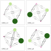
PyEMMA metastable states are represented by colored discs with a radius proportional to their population. The most populated states (labeled 3 and 4) always identify the water phase, while the smaller ones (labeled 1 and 2) represent the two lipid leaflets. Transitions are described with arrows whose thickness and label are associated to the rate constant and MFPT, respectively.
Figure 6
Figure 6
Designation of the kinetic rate constants in membrane planar (A) and spherical (B) geometry. In the liposome model, wo and lo stands for the outer water and lipid compartment, respectively, and similarly for the two inner compartments wi and li.

References

    1. Ju Y., Guo H., Edman M., Hamm-Alvarez S.F. Application of advances in endocytosis and membrane trafficking to drug delivery. Adv. Drug Deliv. Rev. 2020;157:118–141. doi: 10.1016/j.addr.2020.07.026. - DOI - PMC - PubMed
    1. Dean M., Moitra K., Allikmets R. The human ATP-binding cassette (ABC) transporter superfamily. Hum. Mutat. 2022;43:1162–1182. doi: 10.1002/humu.24418. - DOI - PMC - PubMed
    1. Pizzagalli M.D., Bensimon A., Superti-Furga G. A guide to plasma membrane solute carrier proteins. FEBS J. 2021;288:2784–2835. doi: 10.1111/febs.15531. - DOI - PMC - PubMed
    1. Vergalli J., Bodrenko I.V., Masi M., Moynié L., Acosta-Gutiérrez S., Naismith J.H., Davin-Regli A., Ceccarelli M., van den Berg B., Winterhalter M., et al. Porins and small-molecule translocation across the outer membrane of Gram-negative bacteria. Nat. Rev. Microbiol. 2020;18:164–176. doi: 10.1038/s41579-019-0294-2. - DOI - PubMed
    1. Moynié L., Milenkovic S., Mislin G.L.A., Gasser V., Malloci G., Baco E., McCaughan R.P., Page M.G.P., Schalk I.J., Ceccarelli M., et al. The complex of ferric-enterobactin with its transporter from Pseudomonas aeruginosa suggests a two-site model. Nat. Commun. 2019;10:3673. doi: 10.1038/s41467-019-11508-y. - DOI - PMC - PubMed

LinkOut - more resources