

Gene Expression Analysis of Immune Regulatory Genes in Circulating Tumour Cells and Peripheral Blood Mononuclear Cells in Patients with Colorectal Carcinoma
- PMID: 36902476
- PMCID: PMC10003441
- DOI: 10.3390/ijms24055051
Gene Expression Analysis of Immune Regulatory Genes in Circulating Tumour Cells and Peripheral Blood Mononuclear Cells in Patients with Colorectal Carcinoma
Erratum in
-
Correction: Aktar et al. Gene Expression Analysis of Immune Regulatory Genes in Circulating Tumour Cells and Peripheral Blood Mononuclear Cells in Patients with Colorectal Carcinoma. Int. J. Mol. Sci. 2023, 24, 5051.Int J Mol Sci. 2023 Jul 12;24(14):11362. doi: 10.3390/ijms241411362. Int J Mol Sci. 2023. PMID: 37511633 Free PMC article.
Abstract
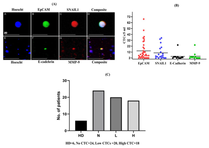
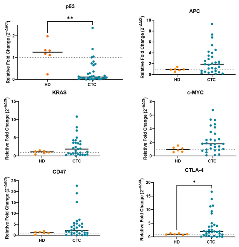
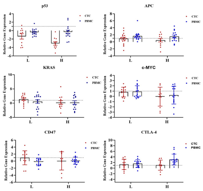
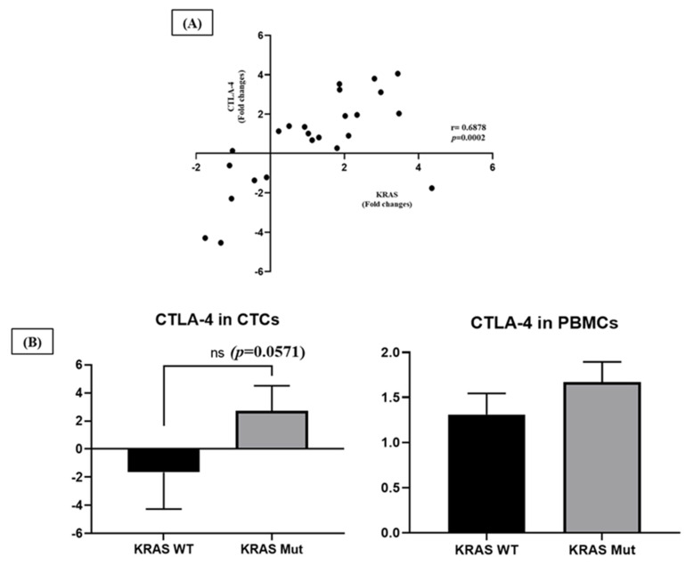
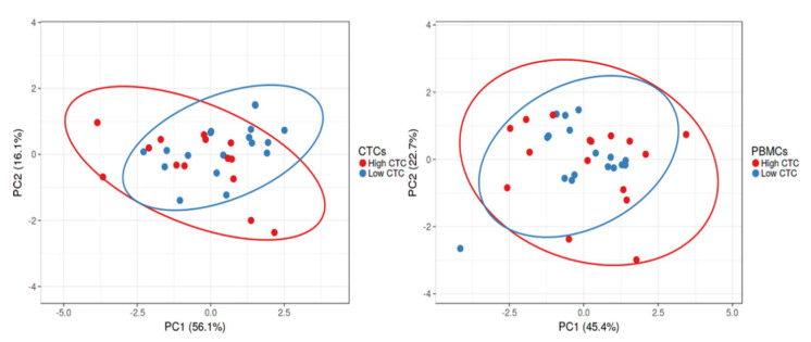
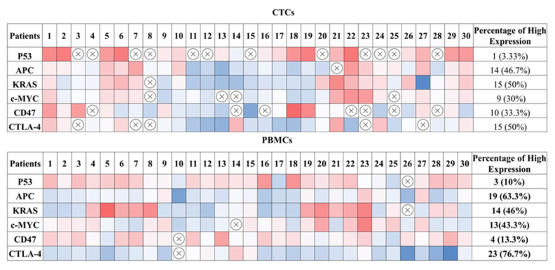
Information regarding genetic alterations of driver cancer genes in circulating tumour cells (CTCs) and their surrounding immune microenvironment nowadays can be employed as a real-time monitoring platform for translational applications such as patient response to therapeutic targets, including immunotherapy. This study aimed to investigate the expression profiling of these genes along with immunotherapeutic target molecules in CTCs and peripheral blood mononuclear cells (PBMCs) in patients with colorectal carcinoma (CRC). Expression of p53, APC, KRAS, c-Myc, and immunotherapeutic target molecules PD-L1, CTLA-4, and CD47 in CTCs and PBMCs were analysed by qPCR. Their expression in high versus low CTC-positive patients with CRC was compared and clinicopathological correlations between these patient groups were analysed. CTCs were detected in 61% (38 of 62) of patients with CRC. The presence of higher numbers of CTCs was significantly correlated with advanced cancer stages (p = 0.045) and the subtypes of adenocarcinoma (conventional vs. mucinous, p = 0.019), while being weakly correlated with tumour size (p = 0.051). Patients with lower numbers of CTCs had higher expression of KRAS. Higher KRAS expression in CTCs was negatively correlated with tumour perforation (p = 0.029), lymph node status (p = 0.037), distant metastasis (p = 0.046) and overall staging (p = 0.004). CTLA-4 was highly expressed in both CTCs and PBMCs. In addition, CTLA-4 expression was positively correlated with KRAS (r = 0.6878, p = 0.002) in the enriched CTC fraction. Dysregulation of KRAS in CTCs might evade the immune system by altering the expression of CTLA-4, providing new insights into the selection of therapeutic targets at the onset of the disease. Monitoring CTCs counts, as well as gene expression profiling of PBMCs, can be helpful in predicting tumour progression, patient outcome and treatment.
Keywords: CTLA-4; KRAS; circulating tumour cells; immune checkpoint molecules; immune escape mechanism; molecular characterisation.
Conflict of interest statement
The authors declare no conflict of interest.
Figures
References
-
- Kallergi G., Vetsika E.-K., Aggouraki D., Lagoudaki E., Koutsopoulos A., Koinis F., Katsarlinos P., Trypaki M., Messaritakis I., Stournaras C., et al. Evaluation of PD-L1/PD-1 on circulating tumor cells in patients with advanced non-small cell lung cancer. Ther. Adv. Med. Oncol. 2018;10:1758834017750121. doi: 10.1177/1758834017750121. - DOI - PMC - PubMed
MeSH terms
Substances
LinkOut - more resources
Full Text Sources
Medical
Research Materials
Miscellaneous
