. 2023 Feb 26;28(5):2183.
doi: 10.3390/molecules28052183.

Molecular Simulation Study on the Interaction between Porcine CR1-like and C3b

Affiliations

Molecular Simulation Study on the Interaction between Porcine CR1-like and C3b

Zhen Hou et al. Molecules. .

Abstract

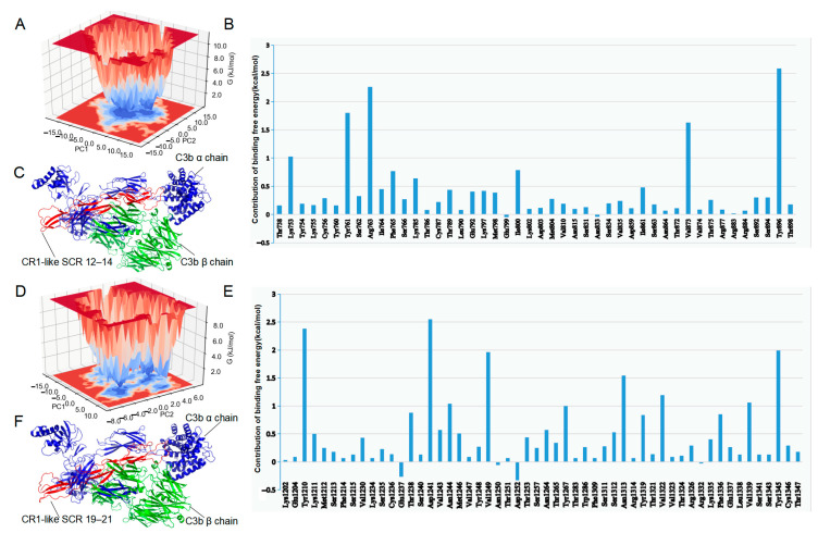
The molecular basis of porcine red blood cell immune adhesion function stems from the complement receptor type 1-like (CR1-like) on its cell membrane. The ligand for CR1-like is C3b, which is produced by the cleavage of complement C3; however, the molecular mechanism of the immune adhesion of porcine erythrocytes is still unclear. Here, homology modeling was used to construct three-dimensional models of C3b and two fragments of CR1-like. An interaction model of C3b-CR1-like was constructed by molecular docking, and molecular structure optimization was achieved using molecular dynamics simulation. A simulated alanine mutation scan revealed that the amino acids Tyr761, Arg763, Phe765, Thr789, and Val873 of CR1-like SCR 12-14 and the amino acid residues Tyr1210, Asn1244, Val1249, Thr1253, Tyr1267, Val1322, and Val1339 of CR1-like SCR 19-21 are key residues involved in the interaction of porcine C3b with CR1-like. This study investigated the interaction between porcine CR1-like and C3b using molecular simulation to clarify the molecular mechanism of the immune adhesion of porcine erythrocytes.

Keywords: C3b; CR1-like; immune adhesion; molecular docking; molecular dynamics.


Conflict of interest statement

The authors declare that the research was conducted in the absence of any commercial or financial relationships that could be construed as potential conflict of interest and competing interests.

Figures

Figure 1
Figure 1
Homology modeling and assessment of C3b and CR1-like fragments. (A) C3b model constructed by AlphaFold-Multimer. The green chain is the C3b β chain, and the cyan chain is the C3b α chain. (B) CR1-like SCR 12–14 model constructed by I-TASSER. The yellow region is the β-fold, and the green region is the loop region. (C) CR1-like SCR 19–21 model constructed by I-TASSER. The yellow region is the β-fold, and the green region is the loop region. (DG) Z scores of C3b β chain, C3b α chain, CR1-like SCR 12–14, and CR1-like SCR 19–21. (HJ) Ramachandran plots of C3b, CR1-like SCR 12–14 and, CR1-like SCR 19–21. Glycine residues are displayed as triangles while other residues are displayed as squares. Residues in the most favored regions (A, B, L) are those with phi and psi angles that are most energetically favorable. Residues in additional allowed regions (a, b, l, p) are less energetically favorable, but still allowed. Residues in generously allowed regions (~a, ~b, ~l, ~p) are the least energetically favorable but still possible.
Figure 2
Figure 2
Molecular dynamics simulation and principal component analysis of CR1-like. (A) Backbone RMSD of CR1-like SCR 12–14 and CR1-like SCR 19–21. (B) Rg of CR1-like SCR 12–14 and CR1-like SCR 19–21. (C,D) PCA-based free energy landscape of CR1-like SCR 12–14 and CR1-like SCR 19–21.
Figure 3
Figure 3
Protein structure optimization of CR1-like fragments. (A,B) CR1-like SCR 12–14 and CR1-like SCR 19–21 models after molecular dynamics simulation. The yellow region is the β-fold, and the green region is the loop region. (C,D) Protein conformational changes of CR1-like SCR 12–14 and CR1-like SCR 19–21 before and after molecular dynamics simulation. Orange models are the conformations before optimization, and blue models are the optimized conformations. (E,F) Ramachandran plots of optimized CR1-like SCR 12–14 and CR1-like SCR 19–21. Glycine residues are displayed as triangles while other residues are displayed as squares. Residues in the most favored regions (A, B, L) are those with phi and psi angles that are most energetically favorable. Residues in additional allowed regions (a, b, l, p) are less energetically favorable, but still allowed. Residues in generously allowed regions (~a, ~b, ~l, ~p) are the least energetically favorable but still possible.
Figure 4
Figure 4
Molecular dynamics simulation of C3b–CR1-like complexes. (A) Backbone RMSD of C3b–CR1-like SCR 12–14 and C3b–CR1-like SCR 19–21. (B) Solvent accessible and surface area of C3b–CR1-like SCR 12–14 and C3b–CR1-like SCR 19–21. (C) Number of hydrogen bonds of C3b–CR1-like SCR 12–14 and C3b–CR1-like SCR 19–21.
Figure 5
Figure 5
Final structure of the complex and simulated alanine mutation scan. (A) PCA-based free energy landscape of the C3b–CR1-like SCR 12–14 complex. (B) Simulated alanine mutation scan of C3b–CR1-like SCR 12–14 complex. (C) Final structure of the C3b–CR1-like SCR 12–14 complex. (D) PCA-based free energy landscape of theC3b–CR1-like SCR 19–21. (E) Simulated alanine mutation scan of C3b–CR1-like SCR 19–21 complex. (F) Final structure of the C3b–CR1-like SCR 19–21 complex.
Figure 6
Figure 6
Prediction and demonstration of CR1-like hotspot residues. (A) Prediction of hotspot residues at the C3b–CR1-like SCR 12–14 binding interface by four prediction tools. (B) Prediction of hotspot residues at the C3b–CR1-like SCR 19–21 binding interface by four prediction tools. (C) Demonstration of C3b–CR1-like SCR 12–14 hotspot residues. (D) Demonstration of C3b–CR1-like SCR 19–21 hotspot residues.

References

    1. Salam K.A., Wang R.Y., Grandinetti T., De Giorgi V., Alter H.J., Allison R.D. Binding of Free and Immune Complex-Associated Hepatitis C Virus to Erythrocytes Is Mediated by the Complement System. Hepatology. 2018;68:2118–2129. doi: 10.1002/hep.30087. - DOI - PMC - PubMed
    1. Anand D., Kumar U., Kanjilal M., Kaur S., Das N. Leucocyte complement receptor 1 (CR1/CD35) transcript and its correlation with the clinical disease activity in rheumatoid arthritis patients. Clin. Exp. Immunol. 2014;176:327–335. doi: 10.1111/cei.12274. - DOI - PMC - PubMed
    1. Chen C.H., Tai S.B., Chen H.C., Yang D.H., Peng M.Y., Lin Y.F. Analysis of Erythrocyte C4d to Complement Receptor 1 Ratio: Use in Distinguishing between Infection and Flare-Up in Febrile Patients with Systemic Lupus Erythematosus. Biomed. Res. Int. 2015;2015:939783. doi: 10.1155/2015/939783. - DOI - PMC - PubMed
    1. Brubaker W.D., Crane A., Johansson J.U. Peripheral complement interactions with amyloid β peptide: Erythrocyte clearance mechanisms. Alzheimers Dement. 2017;13:1397–1409. doi: 10.1016/j.jalz.2017.03.010. - DOI - PMC - PubMed
    1. Khera R., Das N. Complement Receptor 1: Disease associations and therapeutic implications. Mol. Immunol. 2009;46:761–772. doi: 10.1016/j.molimm.2008.09.026. - DOI - PMC - PubMed

Substances

LinkOut - more resources