. 2023 Feb 27;28(5):2195.
doi: 10.3390/molecules28052195.

Baricitinib Attenuates Bleomycin-Induced Pulmonary Fibrosis in Mice by Inhibiting TGF-β1 Signaling Pathway

Affiliations

Baricitinib Attenuates Bleomycin-Induced Pulmonary Fibrosis in Mice by Inhibiting TGF-β1 Signaling Pathway

Songtao Gu et al. Molecules. .

Abstract

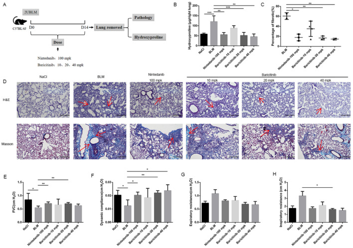
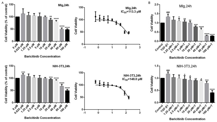
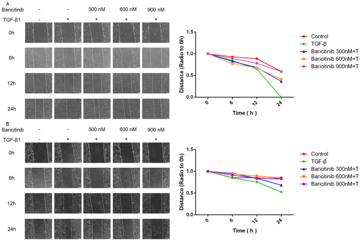
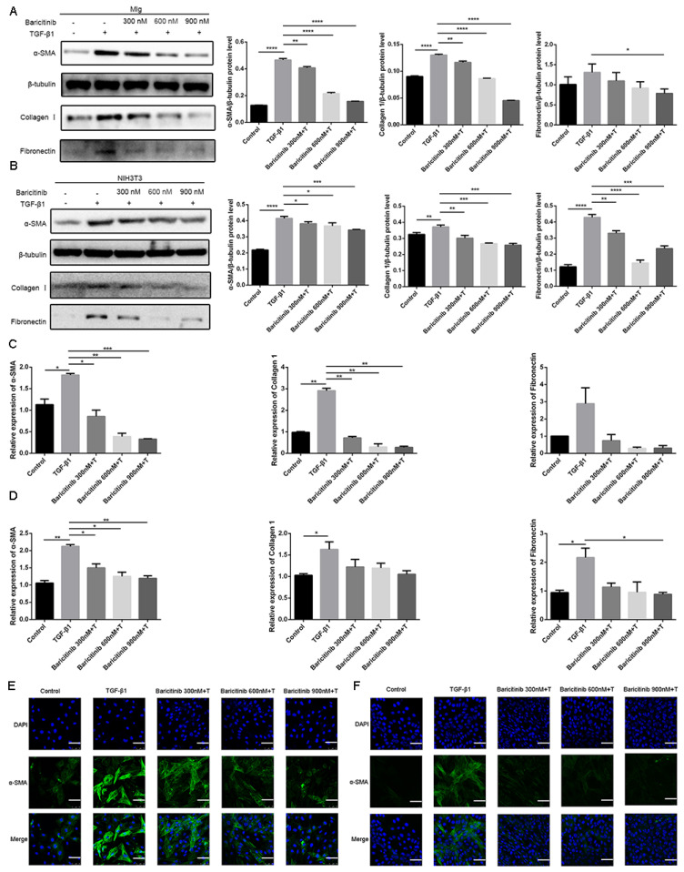
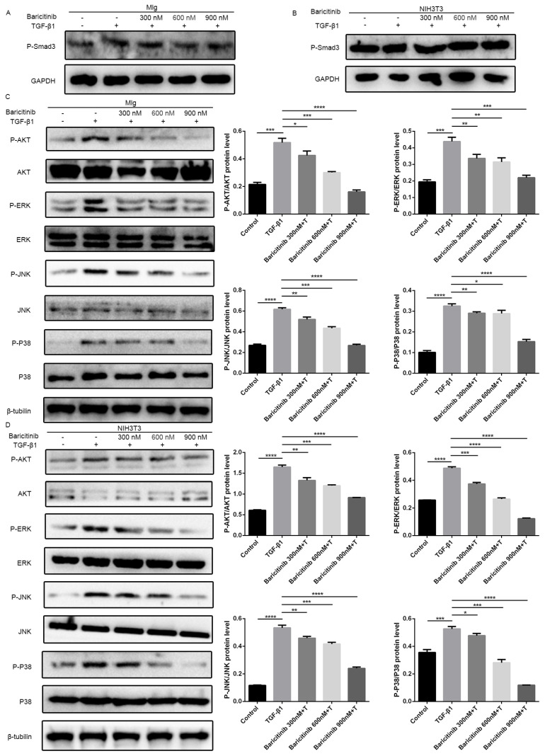
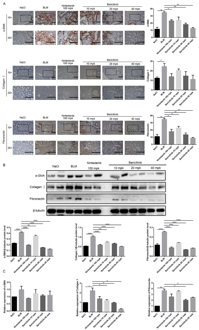
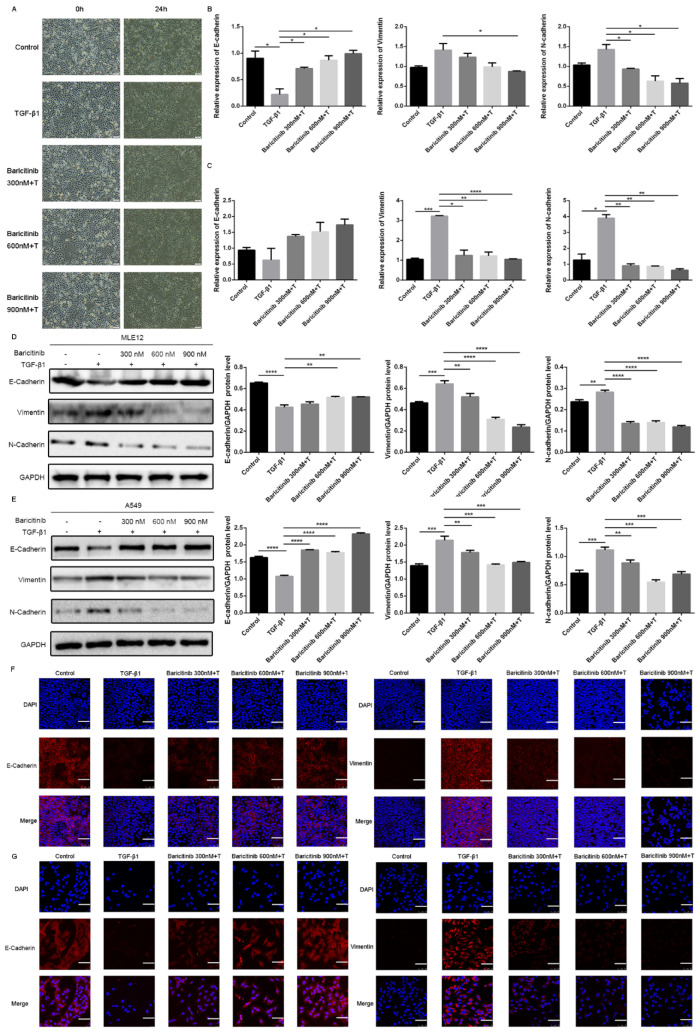
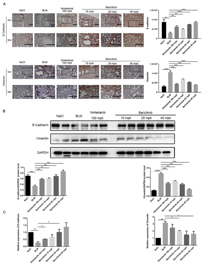
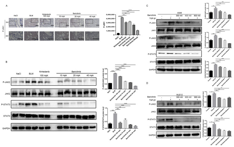
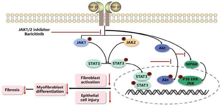
Idiopathic pulmonary fibrosis (IPF) is a chronic progressive interstitial lung disease with unknown etiology, high mortality and limited treatment options. It is characterized by myofibroblast proliferation and extensive deposition of extracellular matrix (ECM), which will lead to fibrous proliferation and the destruction of lung structure. Transforming growth factor-β1 (TGF-β1) is widely recognized as a central pathway of pulmonary fibrosis, and the suppression of TGF-β1 or the TGF-β1-regulated signaling pathway may thus offer potential antifibrotic therapies. JAK-STAT is a downstream signaling pathway regulated by TGF-β1. JAK1/2 inhibitor baricitinib is a marketed drug for the treatment of rheumatoid arthritis, but its role in pulmonary fibrosis has not been reported. This study explored the potential effect and mechanism of baricitinib on pulmonary fibrosis in vivo and in vitro. The in vivo studies have shown that baricitinib can effectively attenuate bleomycin (BLM)-induced pulmonary fibrosis, and in vitro studies showed that baricitinib attenuates TGF-β1-induced fibroblast activation and epithelial cell injury by inhibiting TGF-β1/non-Smad and TGF-β1/JAK/STAT signaling pathways, respectively. In conclusion, baricitinib, a JAK1/2 inhibitor, impedes myofibroblast activation and epithelial injury via targeting the TGF-β1 signaling pathway and reduces BLM-induced pulmonary fibrosis in mice.

Keywords: JAK-STAT; TGF-β1 signaling pathway; baricitinib; pulmonary fibrosis.


Conflict of interest statement

The authors declare no conflict of Interest.

Figures

Figure 1
Figure 1
Baricitinib ameliorates BLM-induced pulmonary fibrosis in mice. (A) Dosing regimen in BLM-induced pulmonary fibrosis model. (B) HYP contents of lung tissues in mice. (C) Statistics of lung fibrosis area among groups. (D) Lung tissue sections were stained with hematoxylin-eosin (HE) and Masson Trichrome staining. Red arrows indicated collagen deposition. (E) Forced vital capacity (FVC) of mice. (F) Dynamic compliance of mice. (G) Expiratory resistance of mice. (H) Inspiratory resistance of mice. Scale bar = 50 μm. Data were noted as the means ± SD, n = 6. * p < 0.05, ** p < 0.01, *** p < 0.001.
Figure 2
Figure 2
Baricitinib suppresses TGF-β1-induced proliferation of fibroblasts. (A) MTT assays of Mlg cells and NIH-3T3 cells. Cells were exposed to the indicated doses of baricitinib (0 to 160 μM) for 24 h, IC50 = 113.3 and 148.0 μM, (n = 5 per group). (B) MTT assays were performed to test the effect of baricitinib on the proliferation of TGF-β1-stimulated Mlg cells and NIH-3T3 cells. Mlg cells and NIH-3T3 cells were treated with baricitinib (0 to 160 μM) and TGF-β1 (5 ng/mL) for 24 h, (n = 5 per group). Data were presented as the means ± SD, n = 5. * represent the difference between TGF-β1-treated group and baricitinib treatment group, # indicated the difference between TGF-β1 treated group versus the control group. * p < 0.05, ** p < 0.01, *** p < 0.001, **** p < 0.0001, ### p < 0.001.
Figure 3
Figure 3
Baricitinib suppresses TGF-β1-induced migration of fibroblasts. (A,B) Wound healing assays of Mlg and NIH-3T3 cells co-cultured with TGF-β1 (5 ng/mL) and baricitinib (300, 600, 900 nM). The wound closure was photographed at 0, 6, 12 and 24 h post-scratching. Data were presented as the means ± SD, n = 3.
Figure 4
Figure 4
Baricitinib attenuates TGF-β1-induced fibroblasts activation. (A,B) Mlg cells and NIH-3T3 cells were treated with TGF-β1 (5 ng/mL) and baricitinib (300, 600, 900 nM) for 24 h. Mlg (A) and NIH-3T3 (B) cells were extracted for Western blot analysis of α-SMA, collagen I and fibronectin. (C,D) Mlg cells and NIH-3T3 cells were treated with TGF-β1 (5 ng/mL) and baricitinib (300, 600, 900 nM) for 24 h. α-SMA (43 kD), β-Tubulin (52 kD) collagen I (220 kD) and fibronectin (260 kD) were analyzed by real-time PCR in Mlg (C) and NIH-3T3 (D) cells. (E,F) Immunofluorescence staining of α-SMA was performed on Mlg (E) and NIH-3T3 (F) cells treated with/without TGF-β1 (5 ng/mL) and/or baricitinib (300, 600, 900 nM) for 24 h. Scale bar = 60 μm, Data were presented as the means ± SD, n = 3. * p < 0.05, ** p < 0.01, *** p < 0.001, **** p < 0.0001.
Figure 5
Figure 5
Baricitinib inhibits TGF-β1-induced activation of non-Smad signaling pathway in fibroblasts. (A,B) Mlg (A) and NIH-3T3 (B) cells were treated with TGF-β1 (5 ng/mL) and baricitinib (300, 600, 900 nM) for 30 min. P-Smad3 (58 kD) were assessed using Western blot. GAPDH (37 kD) was used as the internal control. (C,D) The phosphorylation levels of P-38 (42 kD), JNK (54 kD), ERK (43 kD) and AKT (56 kD) were analyzed by Western blot in Mlg (C) and NIH-3T3 (D) cells treated with TGF-β1 (5 ng/mL) and baricitinib (300, 600, 900 nM) for 1 h. β-tubulin was used as a loading control in grayscale analysis. Data were noted as the means ± SD, n = 3. * p < 0.05, ** p < 0.01, *** p < 0.001, **** p < 0.0001.
Figure 6
Figure 6
Baricitinib inhibits activation of myofibroblasts in vivo. (A) Immunohistochemical staining analysis of α-SMA and collagen I and fibronectin in the lung tissues. Scale bar = 50 μm. (B) Protein levels of α-SMA (43 kD), collagen I (220 kD) and fibronectin (260 kD) were verified by Western blot in lung tissues. β-tubulin (52 kD) was used as an internal reference in densitometric analysis. (C) RT-PCR was performed to detect mRNA levels of α-SMA and collagen I and fibronectin. Data were presented as the means ± SD, n = 3. * p < 0.05, ** p < 0.01, *** p < 0.01, **** p < 0.0001.
Figure 7
Figure 7
Baricitinib inhibits TGF-β1-induced epithelial injury in alveolar epithelial cells. (A) Morphological changes of A549 cells. (B,C) MLE12 and A549 cells were exposed to Baricitinib for 30 min (300, 600, 900 nM) and treated with TGF-β1 (5 ng/mL) for 24 h. (B) mRNA levels E-cadherin, N-cadherin and Vimentin were tested by RT-PCR in MLE12 (B) and A549 (C) cells. (D,E) MLE12 and A549 cells were treated with TGF-β1 (5 ng/mL) and baricitinib (300, 600, 900 nM) for 24 h. Protein expression levels of E-cadherin (120 kD), Vimentin (53 kD), and N-cadherin (100 kD) were assessed by Western blot in A549 (C) and MLE12 (D) cells. GAPDH (37 kD) was used as a loading control. (F,G) MLE12 (F) and A549 (G) cells treated with TGF-β1 (5 ng/mL) and baricitinib (300, 600, 900 nM) for 24 h were immune-stained with E-cadherin and Vimentin. Scale bar = 60 μm. Data were presented as the means ± SD, n = 3. * p < 0.05, ** p < 0.01, *** p < 0.001, **** p < 0.0001.
Figure 8
Figure 8
Baricitinib alleviates epithelial injury in vivo. (A) IHC staining of E-cadherin and Vimentin in the lung tissues. (B) Protein levels of E-cadherin (120 kD), GAPDH (37 kD) and Vimentin (53 kD) in the lung tissues. (C) mRNA expression of E-cadherin and Vimentin in the lung tissues. Scale bar = 50 μm. Data were noted as the means ± SD, n = 3. * p < 0.05; ** p < 0.01; *** p < 0.001; **** p < 0.0001.
Figure 9
Figure 9
Baricitinib alleviates pulmonary fibrosis by inhibiting activation of JAK-STAT signaling pathway. (A) Immunohistochemical staining analysis of P-JAK1 in the lung tissues. Scale bar = 50 μm. (B) The phosphorylation levels of JAK2 and STAT3 were analyzed by Western blotting the lung tissues. (C) The phosphorylation levels of JAK1, JAK2 and STAT3 were analyzed by Western blot in A549 cells treated with TGF-β1 (5 ng/mL) and baricitinib (300, 600, 900 nM) for 1 h. GAPDH (37 kD) was used as a loading control in grayscale analysis. (D) The phosphorylation levels of JAK2 (125 kD) and STAT3 (88 kD) were analyzed by Western blot in MLE12 cells treated with TGF-β1 (5 ng/mL) and baricitinib (300, 600, 900 nM) for 2 h. GAPDH was used as a loading control in grayscale analysis. Data were noted as the means ± SD, n = 3. * p < 0.05; ** p < 0.01; *** p < 0.001; **** p < 0.0001.
Figure 10
Figure 10
Mechanism for the anti-pulmonary fibrosis effect of Baricitinib.

References

    1. Thannickal V.J., Toews G.B., White E.S., Lynch J.P., Martinez F.J. Mechanisms of pulmonary fibrosis. Annu. Rev. Med. 2004;55:395–417. doi: 10.1146/annurev.med.55.091902.103810. - DOI - PubMed
    1. King T.E., Jr., Pardo A., Selman M. Idiopathic pulmonary fibrosis. Lancet. 2011;378:1949–1961. doi: 10.1016/S0140-6736(11)60052-4. - DOI - PubMed
    1. Hardie W.D., Hagood J.S., Davé V., Perl A.-K.T., Whitsett J.A., Korfhagen T.R., Glasser S. Signaling pathways in the epithelial origins of pulmonary fibrosis. Cell Cycle. 2010;9:2769–2776. doi: 10.4161/cc.9.14.12268. - DOI - PMC - PubMed
    1. Meyer K.C. Pulmonary fibrosis, part I: Epidemiology, pathogenesis, and diagnosis. Expert Rev. Respir. Med. 2017;11:343–359. doi: 10.1080/17476348.2017.1312346. - DOI - PubMed
    1. Glass D.S., Grossfeld D., Renna H.A., Agarwala P., Spiegler P., Kasselman L.J., Glass A.D., DeLeon J., Reiss A.B. Idiopathic pulmonary fibrosis: Molecular mechanisms and potential treatment approaches. Respir. Investig. 2020;58:320–335. doi: 10.1016/j.resinv.2020.04.002. - DOI - PubMed