

Superparamagnetic Iron-Oxide Nanoparticles Synthesized via Green Chemistry for the Potential Treatment of Breast Cancer
- PMID: 36903587
- PMCID: PMC10005561
- DOI: 10.3390/molecules28052343
Superparamagnetic Iron-Oxide Nanoparticles Synthesized via Green Chemistry for the Potential Treatment of Breast Cancer
Abstract
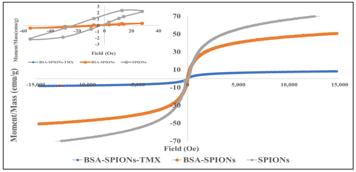
In the emerging field of nanomedicine, nanoparticles have been widely considered as drug carriers and are now used in various clinically approved products. Therefore, in this study, we synthesized superparamagnetic iron-oxide nanoparticles (SPIONs) via green chemistry, and the SPIONs were further coated with tamoxifen-conjugated bovine serum albumin (BSA-SPIONs-TMX). The BSA-SPIONs-TMX were within the nanometric hydrodynamic size (117 ± 4 nm), with a small poly dispersity index (0.28 ± 0.02) and zeta potential of -30.2 ± 0.09 mV. FTIR, DSC, X-RD, and elemental analysis confirmed that BSA-SPIONs-TMX were successfully prepared. The saturation magnetization (Ms) of BSA-SPIONs-TMX was found to be ~8.31 emu/g, indicating that BSA-SPIONs-TMX possess superparamagnetic properties for theragnostic applications. In addition, BSA-SPIONs-TMX were efficiently internalized into breast cancer cell lines (MCF-7 and T47D) and were effective in reducing cell proliferation of breast cancer cells, with IC50 values of 4.97 ± 0.42 μM and 6.29 ± 0.21 μM in MCF-7 and T47D cells, respectively. Furthermore, an acute toxicity study on rats confirmed that these BSA-SPIONs-TMX are safe for use in drug delivery systems. In conclusion, green synthesized superparamagnetic iron-oxide nanoparticles have the potential to be used as drug delivery carriers and may also have diagnostic applications.
Keywords: SPIONs; breast cancer; green chemistry; serum albumin; superparamagnetic iron-oxide nanoparticles; tamoxifen.
Conflict of interest statement
The authors declare no conflict of interest.
Figures


Similar articles
-
Superparamagnetic iron oxide nanoparticles conjugated with folic acid for dual target-specific drug delivery and MRI in cancer theranostics.Mater Sci Eng C Mater Biol Appl. 2017 Jan 1;70(Pt 1):763-771. doi: 10.1016/j.msec.2016.09.052. Epub 2016 Sep 26. Mater Sci Eng C Mater Biol Appl. 2017. PMID: 27770953
-
Heparin-Superparamagnetic Iron Oxide Nanoparticles for Theranostic Applications.Molecules. 2022 Oct 21;27(20):7116. doi: 10.3390/molecules27207116. Molecules. 2022. PMID: 36296711 Free PMC article.
-
Polyethylene Glycol-Chitosan Oligosaccharide-Coated Superparamagnetic Iron Oxide Nanoparticles: A Novel Drug Delivery System for Curcumin Diglutaric Acid.Biomolecules. 2020 Jan 2;10(1):73. doi: 10.3390/biom10010073. Biomolecules. 2020. PMID: 31906490 Free PMC article.
-
Superparamagnetic iron oxide nanoparticles: magnetic nanoplatforms as drug carriers.Int J Nanomedicine. 2012;7:3445-71. doi: 10.2147/IJN.S30320. Epub 2012 Jul 6. Int J Nanomedicine. 2012. PMID: 22848170 Free PMC article. Review.
-
Superparamagnetic iron oxide nanoparticles for delivery of therapeutic agents: opportunities and challenges.Expert Opin Drug Deliv. 2014 Sep;11(9):1449-70. doi: 10.1517/17425247.2014.924501. Epub 2014 May 29. Expert Opin Drug Deliv. 2014. PMID: 24870351 Review.
Cited by
-
Synthesis of Quercetin-Loaded Silver Nanoparticles and Assessing Their Anti-Bacterial Potential.Micromachines (Basel). 2023 Nov 25;14(12):2154. doi: 10.3390/mi14122154. Micromachines (Basel). 2023. PMID: 38138323 Free PMC article.
-
Updates on Biogenic Metallic and Metal Oxide Nanoparticles: Therapy, Drug Delivery and Cytotoxicity.Pharmaceutics. 2023 Jun 3;15(6):1650. doi: 10.3390/pharmaceutics15061650. Pharmaceutics. 2023. PMID: 37376098 Free PMC article. Review.
-
A Comprehensive Review of Nanostructured Lipid Carriers: Innovations and Applications in Breast Cancer Treatment.Recent Adv Drug Deliv Formul. 2025;19(1):25-44. doi: 10.2174/0126673878313086241031154146. Recent Adv Drug Deliv Formul. 2025. PMID: 39660493 Review.
-
Mannose-Functionalized Isoniazid-Loaded Nanostructured Lipid Carriers for Pulmonary Delivery: In Vitro Prospects and In Vivo Therapeutic Efficacy Assessment.Pharmaceuticals (Basel). 2023 Aug 4;16(8):1108. doi: 10.3390/ph16081108. Pharmaceuticals (Basel). 2023. PMID: 37631023 Free PMC article.
-
Roadmap on magnetic nanoparticles in nanomedicine.Nanotechnology. 2024 Nov 5;36(4):042003. doi: 10.1088/1361-6528/ad8626. Nanotechnology. 2024. PMID: 39395441 Free PMC article. Review.
References
MeSH terms
Substances
Grants and funding
LinkOut - more resources
Full Text Sources
Medical
Miscellaneous
