. 2023 Mar 3;12(5):1167.
doi: 10.3390/plants12051167.

Antiphotoaging and Skin-Protective Activities of Ardisia silvestris Ethanol Extract in Human Keratinocytes

Affiliations

Antiphotoaging and Skin-Protective Activities of Ardisia silvestris Ethanol Extract in Human Keratinocytes

Lei Huang et al. Plants (Basel). .

Abstract

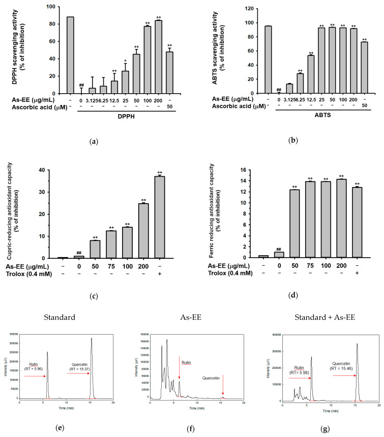
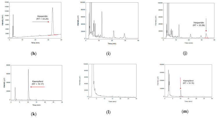
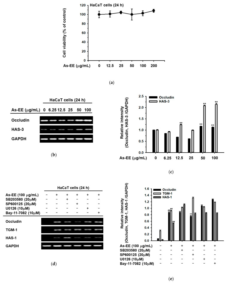
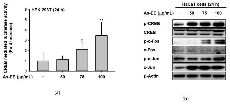
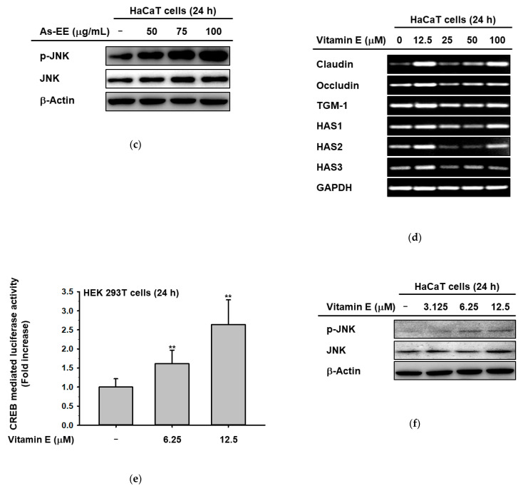
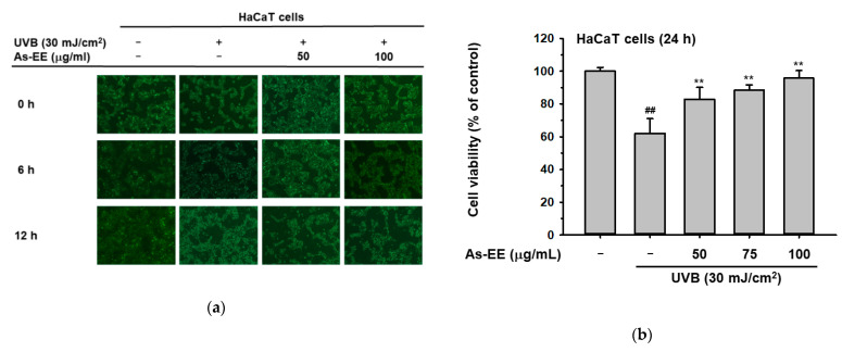
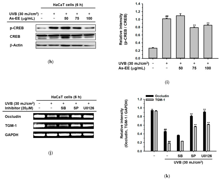
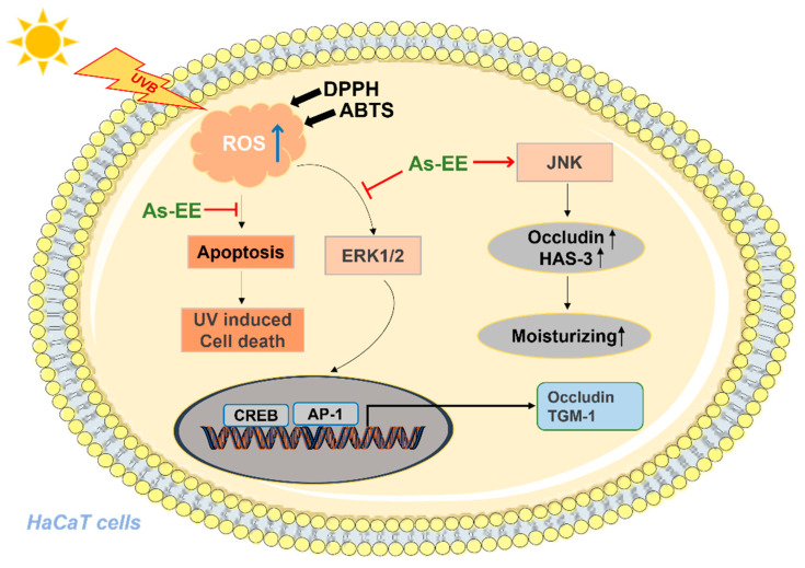
Ardisia silvestris is a traditional medicinal herb used in Vietnam and several other countries. However, the skin-protective properties of A. silvestris ethanol extract (As-EE) have not been evaluated. Human keratinocytes form the outermost barrier of the skin and are the main target of ultraviolet (UV) radiation. UV exposure causes skin photoaging via the production of reactive oxygen species. Protection from photoaging is thus a key component of dermatological and cosmetic products. In this research, we found that As-EE can prevent UV-induced skin aging and cell death as well as enhance the barrier effect of the skin. First, the radical-scavenging ability of As-EE was checked using DPPH, ABTS, TPC, CUPRAC, and FRAP assays, and a 3-(4-5-dimethylthiazol-2-yl)-2-5-diphenyltetrazolium bromide assay was used to examine cytotoxicity. Reporter gene assays were used to determine the doses that affect skin-barrier-related genes. A luciferase assay was used to identify possible transcription factors. The anti-photoaging mechanism of As-EE was investigated by determining correlated signaling pathways using immunoblotting analyses. As-EE had no harmful effects on HaCaT cells, according to our findings, and As-EE revealed moderate radical-scavenging ability. With high-performance liquid chromatography (HPLC) analysis, rutin was found to be one of the major components. In addition, As-EE enhanced the expression levels of hyaluronic acid synthase-1 and occludin in HaCaT cells. Moreover, As-EE dose-dependently up-regulated the production of occludin and transglutaminase-1 after suppression caused by UVB blocking the activator protein-1 signaling pathway, in particular, the extracellular response kinase and c-Jun N-terminal kinase. Our findings suggest that As-EE may have anti-photoaging effects by regulating mitogen-activated protein kinase, which is good news for the cosmetics and dermatology sectors.

Keywords: AP-1; Ardisia silvestris ethanol extract; ROS; UVB irradiation; anti-apoptosis; antioxidative capacity.


Conflict of interest statement

The authors have no conflict of interest to declare.

Figures

Figure 1
Figure 1
Effects of As-EE on antioxidative capacity. (a) As-EE (0 to 200 µg/mL) was incubated with DPPH (250 µM) in foil at 37 °C for 30 min and then the absorbance was measured at 517 nm. Ascorbic acid (50 µM) together with positive controls; (b) ABTS and potassium persulfate solution mixed with As-EE (0 to 200 µg/mL) incubated for 30 min in the dark at 37 °C. At 730 nm, the solution’s absorbance was found; (c) Copper(II) chloride solution, neocuproine solution, and NH4Ac buffer were mixed with As-EE (0 to 200 µg/mL). Trolox (0.4 mM) was used as a positive control. After 1 h of incubation, the absorbance was measured at 450 nm using a spectrophotometer; (d) Acetic acid buffer, TPTZ solution, and FeCl3 solution were mixed with As-EE (0 to 200 µg/mL). A positive control was employed, which was trolox (0.4 mM). Each fraction’s absorbance was measured at 593 nm after 15 min of dark incubation at 37 °C; (em) The phytochemical profiles of rutin, quercetin, hesperidin, and kaempferol in As-EE were analyzed using HPLC. Results (ad) are expressed as the mean ± standard deviation. # p < 0.05 and ## p < 0.01 compared with the normal groups. * p < 0.05 and ** p < 0.01 compared with the control groups.
Figure 1
Figure 1
Effects of As-EE on antioxidative capacity. (a) As-EE (0 to 200 µg/mL) was incubated with DPPH (250 µM) in foil at 37 °C for 30 min and then the absorbance was measured at 517 nm. Ascorbic acid (50 µM) together with positive controls; (b) ABTS and potassium persulfate solution mixed with As-EE (0 to 200 µg/mL) incubated for 30 min in the dark at 37 °C. At 730 nm, the solution’s absorbance was found; (c) Copper(II) chloride solution, neocuproine solution, and NH4Ac buffer were mixed with As-EE (0 to 200 µg/mL). Trolox (0.4 mM) was used as a positive control. After 1 h of incubation, the absorbance was measured at 450 nm using a spectrophotometer; (d) Acetic acid buffer, TPTZ solution, and FeCl3 solution were mixed with As-EE (0 to 200 µg/mL). A positive control was employed, which was trolox (0.4 mM). Each fraction’s absorbance was measured at 593 nm after 15 min of dark incubation at 37 °C; (em) The phytochemical profiles of rutin, quercetin, hesperidin, and kaempferol in As-EE were analyzed using HPLC. Results (ad) are expressed as the mean ± standard deviation. # p < 0.05 and ## p < 0.01 compared with the normal groups. * p < 0.05 and ** p < 0.01 compared with the control groups.
Figure 2
Figure 2
Effects of As-EE on cell viability and skin moisture-protection activity. (a) Cytotoxicity of As-EE was measured using a 3-(4-5-dimethylthiazol-2-yl)-2,5-diphenyltetrazolium bromide (MTT) assay in HaCaT cells; (b) The expression levels of skin-barrier-related and hydration factors were determined with RT-PCR in HaCaT cells after treatment with As-EE at doses ranging from 0 to 100 µg/mL for 24 h; (c) The relative intensity of RT-PCR results (occludin and HAS-3) was measured with ImageJ; (d) Inhibitors were applied to HaCaT cells (SB20580, a p38 inhibitor; SP600125, a JNK inhibitor; U0126, an ERK inhibitor; and Bay117082, a κB kinase inhibitor) for 24 h and the mRNA levels of occludin, TGM-1, and HAS-1 were measured with RT-PCR. (e) The relative intensity of mRNA levels was measured with ImageJ. # p < 0.05 and ## p < 0.01 compared with the normal groups. * p < 0.05 and ** p < 0.01 compared with the control groups.
Figure 3
Figure 3
Effects of As-EE on MAPK-mediated AP-1 and CREB signaling pathway. (a) HEK293T cells transfected with CREB-Luc were incubated with As-EE for 24 h. A galactosidase construct was used as a control, and luciferase activity was measured using a luminometer; (b,c) HaCaT cells were incubated with As-EE for 24 h. Immunoblot analysis was used to evaluate the phosphorylation levels of c-Jun, c-Fos, p-CREB, and JNK; (d) RT-PCR was used to evaluate the expression levels of skin-barrier-related and hydration factors in HaCaT cells after treatment with vitamin E in a dose-dependent manner from 0 to 100 µM for 24 h; (e) Luciferase assay; (f) Phosphorylation level of JNK was measured with immunoblotting in HaCaT cells treated with vitamin E for 24 h. * p < 0.05 and ** p < 0.01 compared with the normal groups.
Figure 3
Figure 3
Effects of As-EE on MAPK-mediated AP-1 and CREB signaling pathway. (a) HEK293T cells transfected with CREB-Luc were incubated with As-EE for 24 h. A galactosidase construct was used as a control, and luciferase activity was measured using a luminometer; (b,c) HaCaT cells were incubated with As-EE for 24 h. Immunoblot analysis was used to evaluate the phosphorylation levels of c-Jun, c-Fos, p-CREB, and JNK; (d) RT-PCR was used to evaluate the expression levels of skin-barrier-related and hydration factors in HaCaT cells after treatment with vitamin E in a dose-dependent manner from 0 to 100 µM for 24 h; (e) Luciferase assay; (f) Phosphorylation level of JNK was measured with immunoblotting in HaCaT cells treated with vitamin E for 24 h. * p < 0.05 and ** p < 0.01 compared with the normal groups.
Figure 4
Figure 4
Moisturizing and anti-apoptotic effect of As-EE in UVB-irradiated human keratinocytes. (a) The morphology of HaCaT cells with As-EE treatment (50 and 100 µg/mL) under UVB irradiation for 6 and 12 h was examined using microscopy; (b) Viability of HaCaT cells was evaluated with an MTT assay in As-EE-treated cells exposed to UVB irradiation; (c) FACS analysis in HaCaT cells treated with As-EE under UVB irradiation; (d) HaCaT cells were pre-treated with As-EE for 30 min and irradiated with UVB. After incubation for 6 h, the mRNA levels were measured with RT-PCR; (e) The relative intensity of mRNA was measured with ImageJ; (f) Phosphorylation levels of ERK were checked with an immunoblot analysis; (g) The relative intensity of protein was measured using ImageJ; (h) Phosphorylation levels of CREB were checked with an immunoblot analysis; (i) The relative intensity of protein was measured using ImageJ; (j) HaCaT cells were treated with MAPK inhibitors (SB20580, a p38 inhibitor; SP600125, a JNK inhibitor; and U0126, an ERK inhibitor) for 6 h under UVB irradiation and mRNA levels were determined with RT-PCR; (k) The relative intensity of mRNA levels was measured with ImageJ. * p < 0.05 and ** p < 0.01 compared with the control groups (only UVB group). # p < 0.05 and ## p < 0.01 compared with the normal groups.
Figure 4
Figure 4
Moisturizing and anti-apoptotic effect of As-EE in UVB-irradiated human keratinocytes. (a) The morphology of HaCaT cells with As-EE treatment (50 and 100 µg/mL) under UVB irradiation for 6 and 12 h was examined using microscopy; (b) Viability of HaCaT cells was evaluated with an MTT assay in As-EE-treated cells exposed to UVB irradiation; (c) FACS analysis in HaCaT cells treated with As-EE under UVB irradiation; (d) HaCaT cells were pre-treated with As-EE for 30 min and irradiated with UVB. After incubation for 6 h, the mRNA levels were measured with RT-PCR; (e) The relative intensity of mRNA was measured with ImageJ; (f) Phosphorylation levels of ERK were checked with an immunoblot analysis; (g) The relative intensity of protein was measured using ImageJ; (h) Phosphorylation levels of CREB were checked with an immunoblot analysis; (i) The relative intensity of protein was measured using ImageJ; (j) HaCaT cells were treated with MAPK inhibitors (SB20580, a p38 inhibitor; SP600125, a JNK inhibitor; and U0126, an ERK inhibitor) for 6 h under UVB irradiation and mRNA levels were determined with RT-PCR; (k) The relative intensity of mRNA levels was measured with ImageJ. * p < 0.05 and ** p < 0.01 compared with the control groups (only UVB group). # p < 0.05 and ## p < 0.01 compared with the normal groups.
Figure 5
Figure 5
Summary of pathways regulated by As-EE related to its moisturizing and anti-photoaging effects.

Similar articles

Cited by

References

    1. You L., Cho J.Y. The Regulatory Role of Korean Ginseng in Skin Cells. J. Ginseng Res. 2021;45:363–370. doi: 10.1016/j.jgr.2020.08.004. - DOI - PMC - PubMed
    1. Chambers E.S., Vukmanovic-Stejic M. Skin Barrier Immunity and Ageing. Immunology. 2020;160:116–125. doi: 10.1111/imm.13152. - DOI - PMC - PubMed
    1. Than U.T.T., Leavesley D.I., Parker T.J. Characteristics and Roles of Extracellular Vesicles Released by Epidermal Keratinocytes. J. Eur. Acad. Dermatol. Venereol. 2019;33:2264–2272. doi: 10.1111/jdv.15859. - DOI - PubMed
    1. Rawlings A.V., Harding C.R. Moisturization and Skin Barrier Function. Dermatol. Ther. 2004;17:43–48. doi: 10.1111/j.1396-0296.2004.04S1005.x. - DOI - PubMed
    1. Baroni A., Buommino E., De Gregorio V., Ruocco E., Ruocco V., Wolf R. Structure and Function of the Epidermis Related to Barrier Properties. Clin. Dermatol. 2012;30:257–262. doi: 10.1016/j.clindermatol.2011.08.007. - DOI - PubMed