. 2023 Feb 22;15(5):1090.
doi: 10.3390/nu15051090.

Biomimetic Nanozymes Suppressed Ferroptosis to Ameliorate Doxorubicin-Induced Cardiotoxicity via Synergetic Effect of Antioxidant Stress and GPX4 Restoration

Affiliations

Biomimetic Nanozymes Suppressed Ferroptosis to Ameliorate Doxorubicin-Induced Cardiotoxicity via Synergetic Effect of Antioxidant Stress and GPX4 Restoration

Yunpeng Zhang et al. Nutrients. .

Abstract

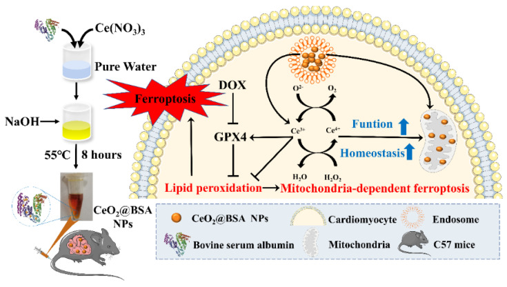
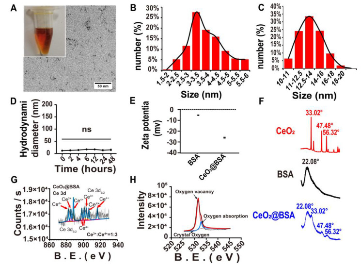
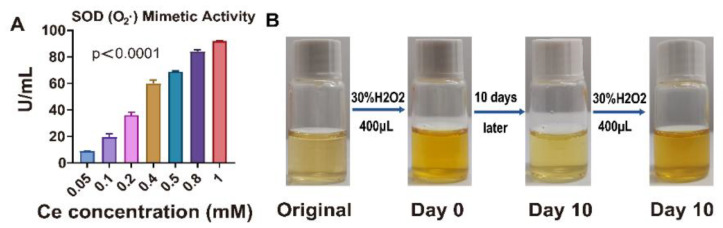
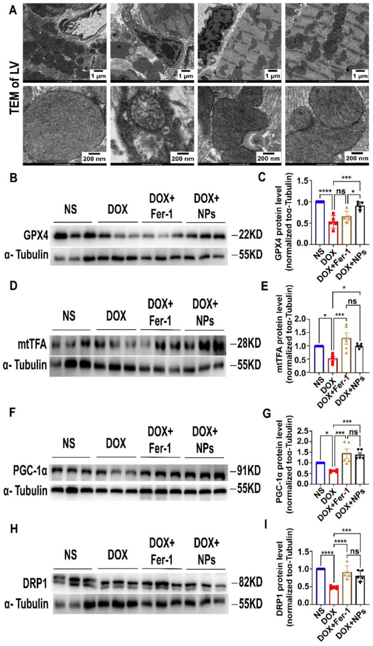
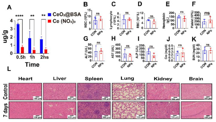
Mitochondria-dependent ferroptosis plays an important role in the pathogenesis of doxorubicin (DOX)-induced cardiotoxicity (DIC), which remains a clinical challenge due to the lack of effective interventions. Cerium oxide (CeO2), a representative nanozyme, has attracted much attention because of its antioxidant properties. This study evaluated CeO2-based nanozymes for the prevention and treatment of DIC in vitro and in vivo by adding nanoparticles (NPs), which were synthesized by biomineralization, to the culture or giving them to the mice, and the ferroptosis-specific inhibitor ferrostatin-1 (Fer-1) was used as control. The prepared NPs exhibited an excellent antioxidant response and glutathione peroxidase 4 (GPX4)-depended bioregulation, with the additional merits of bio-clearance and long retention in the heart. The experiments showed that NP treatment could significantly reverse myocardial structural and electrical remodeling, and reduce myocardial necrosis. These cardioprotective therapeutic effects were associated with their ability to alleviate oxidative stress, mitochondrial lipid peroxidation, and mitochondrial membrane potential damage, with a superior efficiency to the Fer-1. The study also found that the NPs significantly restored the expression of GPX4 and mitochondrial-associated proteins, thereby restoring mitochondria-dependent ferroptosis. Therefore, the study provides some insights into the role of ferroptosis in DIC. It also shows that CeO2-based nanozymes could be a promising prevention and treatment candidate as a novel cardiomyocyte ferroptosis protector to mitigate DIC and improve prognosis and quality of life in cancer patients.

Keywords: biomineralization; doxorubicin-induced cardiomyopathy; ferroptosis; mitochondria; nanozyme; oxidative stress.


Conflict of interest statement

The authors declare no conflict of interest.

Figures

Figure 1
Figure 1
Scheme of biomimetic cerium-oxide-based nanozyme synthesis by biomineralization. After tail vein injection, the nanozymes were taken up by the cardiomyocytes and mitochondria, activating the GPX4 to reverse ferroptosis by maintaining mitochondrial function and homeostasis. BSA: bovine serum protein, NPs: nanoparticles, DOX: doxorubicin, GPX4: glutathione peroxidase 4.
Figure 2
Figure 2
CeO2@BSA NP characterization. (A) Appearance and TEM images (Scale bar = 50 nm) of NPs. (B) Size distribution of nanoparticles as shown in (A). (C) Diameter distribution of nanoparticles measured via DLS. (D) Particle size at different points in 48 h measured by DLS. (E) Zeta potential of BSA and NPs. (F) XRD of CeO2, BSA, and NPs. (G) XPS of the spectrum of the Ce3d. (H) XPS of the spectrum of the O1s. TEM: transmission electron microscopy, DLS: dynamic light scattering, XRD: X−ray diffraction, XPS: X−ray photoelectron spectroscopy. Data are expressed as mean ± SEM of three independent replicates.
Figure 3
Figure 3
Superoxide dismutase (SOD)- and catalase (CAT)-hybrid-mimetic enzyme activities of CeO2@BSA NPs in vitro. (A) Statistical graphs of SOD-mimetic enzyme activity of CeO2@BSA NPs. (B) The digital images showed the coloration of CeO2@BSA NP solutions moments after the addition of H2O2. Ce3+ redox state is peculiar to colorless ions and the coloration is peculiar to the Ce4+ redox state. A change in color indicates a change in Ce4+/Ce3+. H2O2: hydrogen peroxide. The data are expressed as mean ± SEM of three independent replicates.
Figure 4
Figure 4
CeO2@BSA NPs protect against DIC by preventing ferroptosis in H9c2 cell lines. (A) MTT cell viability experiment of the different concentrations of CeO2@BSA NPs measured by cerium concentration on H9c2 cells at 24 h. (B) Representative confocal microscopy images of the cellular uptake of CeO2@BSA NPs at 6hrs, scale bar = 100 μm. (C) Fluorescent images of H9c2 cells that were stained with DCFH-DA, scale bar = 50 μm. (D) Representative confocal microscopy images of H9c2 cells that were stained with JC-1 dye, scale bar = 50 μm. (E) Representative fluorescent images of H9c2 cells’ mitochondrial ferrous ions that were stained with Mito-FerroGreen, scale bar = 100 μm. (F) Representative fluorescent images of H9c2 cells’ mitochondrial lipid peroxides that were stained with MitoPeDPP, scale bar = 100 μm. (G) Quantification of ROS via DCFH-DA intensity in (C). (H) Quantification of MMP of H9c2 cells via JC-1 monomer/JC-1 aggregates in (D). (I) Quantification of ferrous ions in mitochondria measured by Mito-FerroGreen intensity in (E). (J) Quantification of mitochondrial lipid peroxides by MitoPeDPP intensity in (F). DAPI: 4′,6′-diamidino-2-phenylindole, FITC: fluorescein isothiocyanate, DOX: doxorubicin, Fer-1: ferrostatin-1, NPs: CeO2@BSA nanoparticles, PBS: phosphate buffer solution, H2O2: hydrogen peroxide; DCFH-DA: 2′,7′-dichlorofluorescin diacetate; ROS: reactive oxygen species; MMP: mitochondrial membrane potential. Data are expressed as mean ± SEM of three to five independent replicates; * p < 0.05, ** p < 0.01, *** p < 0.001, **** p < 0.001, ns: no statistical difference.
Figure 5
Figure 5
Therapeutic effects of CeO2@BSA NPs on doxorubicin-induced cardiac dysfunction in mice. (A) Schematic animal experiment and intervention protocol. (B) Statistical chart of weight change in four groups. (C) Analysis results of LVEF in M-mode of echocardiographic images. (D) Analysis results of LVFS in M-mode of echocardiographic images. (E) Analysis results of LVID in M-mode of echocardiographic images. (F) Analysis results of LV Vol in (C). (G) Analysis results of PR interval of electrocardiography. (H) Statistical chart of serum cTnI in four groups. (I) Statistical chart of serum NT-proBNP in four groups. (J) Representative epicardial electrical mapping recorded for LV. (K) Conduction velocity of LV in (J). (L) Absolute inhomogeneity of LV in (J). (M) Inhomogeneity index of LV in (J). (N) Statistical chart of serum GSH in four groups. (O) Statistical chart of serum MDA in four groups. (P) Representative HE staining of myocardial tissue in four groups, scale bar = 50 μm or 20 μm. NS: normal saline, DOX: doxorubicin, Fer-1: ferrostatin-1, NPs: CeO2@BSA nanoparticles, LV: left ventricle, LVEF: left ventricular ejection fraction, LVFS: left ventricular fractional shortening, LVID: left ventricular internal diameter, LV Vol: left ventricular volume, cTnI: cardiac troponin I, NT-proBNP: N-terminal brain natriuretic peptide, GSH: glutathione, MDA: malondialdehyde, HE staining: Hematoxylin-eosin staining. Data are expressed as mean ± SEM of three to six independent replicates; * p < 0.05, ** p < 0.01, *** p < 0.001, **** p < 0.001, ns: no statistical difference.
Figure 6
Figure 6
CeO2@BSA NPs exhibit a protective effect against ferroptosis by maintaining mitochondrial morphology, increasing GPX4, and mitochondria-related protein expression. (A) TEM images of mitochondria in mice cardiomyocytes, scale bar = 1 μm or 200 nm. (B) Representative Western blot results of GPX4 in the four groups. (C) Analysis of the protein expression of GPX4 in the four groups. (D) Representative Western blot results of mtTFA in the four groups. (E) Analysis of the protein expression of mtTFA in the four groups. (F) Representative Western blot results of PGC-1α in the four groups. (G) Analysis of the protein expression of PGC-1α in the four groups. (H) Representative Western blot results of DRP1 in the four groups. (I) Analysis of the protein expression of DRP1 in the four groups. NS: normal saline, DOX: doxorubicin, Fer-1: ferro-statin-1, NPs: CeO2@BSA nanoparticles, GPX4: glutathione peroxidase 4, mtTFA: mitochondrial transcription factor A, PGC-1α: α subunit of peroxisome proliferators-activated receptor-γcoactivator-1, DRP1: dynamin-related protein 1. Data are expressed as mean ± SEM of three to five independent replicates; * p < 0.05, *** p < 0.001, **** p < 0.001, ns: no statistical difference.
Figure 7
Figure 7
In vivo metabolism and biocompatibility of CeO2@BSA NPs. (A) Cardiac uptake of CeO2@BSA NPs and the equivalent cerium content of Ce(NO3)3 at different time points after tail vein injection. (BF) Analysis of the blood routine examination of WBC (B), lymphocytes (C), RBC (D), hemoglobin (E), and platelets (F) in the two groups. (GI). Analysis of the liver function via ALT (G), AST (H), and ALP (I) in the two groups. (J,K) Analysis of the renal function via Cre (J) and BUN (K) in the two groups. (L). Representative HE staining of heart, liver, spleen, lung, kidney, and brain in the two groups after a week of treatment, scale bar = 100 μm. WBC: white blood cell, RBC: red blood cell, ALT: alanine transaminase, AST: aspartate transaminase, ALP: alkaline phosphatase, Cre: creatinine, BUN: blood urea nitrogen, HE staining: hematoxylin/eosin (HE). Data are expressed as mean ± SEM of three independent replicates; ** p < 0.01, **** p < 0.001, ns: no statistical difference.

Similar articles

Cited by

References

    1. Armenian S., Xu L., Ky B., Sun C., Farol L., Pal S., Douglas P., Bhatia S., Chao C. Cardiovascular Disease Among Survivors of Adult-Onset Cancer: A Community-Based Retrospective Cohort Study. J. Clin. Oncol. 2016;34:1122–1130. doi: 10.1200/JCO.2015.64.0409. - DOI - PMC - PubMed
    1. Lipshultz S., Adams M., Colan S., Constine L., Herman E., Hsu D., Hudson M., Kremer L., Landy D., Miller T., et al. Long-term cardiovascular toxicity in children, adolescents, and young adults who receive cancer therapy: Pathophysiology, course, monitoring, management, prevention, and research directions: A scientific statement from the American Heart Association. Circulation. 2013;128:1927–1995. doi: 10.1161/CIR.0b013e3182a88099. - DOI - PubMed
    1. Rugbjerg K., Mellemkjaer L., Boice J., Køber L., Ewertz M., Olsen J. Cardiovascular disease in survivors of adolescent and young adult cancer: A Danish cohort study, 1943–2009. J. Natl. Cancer Inst. 2014;106:dju110. doi: 10.1093/jnci/dju110. - DOI - PubMed
    1. Swain S., Whaley F., Ewer M. Congestive heart failure in patients treated with doxorubicin: A retrospective analysis of three trials. Cancer. 2003;97:2869–2879. doi: 10.1002/cncr.11407. - DOI - PubMed
    1. Nabhan C., Byrtek M., Rai A., Dawson K., Zhou X., Link B., Friedberg J., Zelenetz A., Maurer M., Cerhan J., et al. Disease characteristics, treatment patterns, prognosis, outcomes and lymphoma-related mortality in elderly follicular lymphoma in the United States. Br. J. Haematol. 2015;170:85–95. doi: 10.1111/bjh.13399. - DOI - PMC - PubMed